Japanese
English
- 有料閲覧
PPV購入案内
1,320円 (1,200円+税10%) こちらは、195ページから198ページの文献です。購入には医書.jp本会員登録が必要です。
会員登録完了後に改めて上記「購入サイトへ」を選択してください。
または「購入サイトへ」選択後に「購入ログイン」、「新規会員登録」の順に進んでください。
会員登録について詳しくはをご確認ください。
- Abstract 文献概要
- 1ページ目 Look Inside
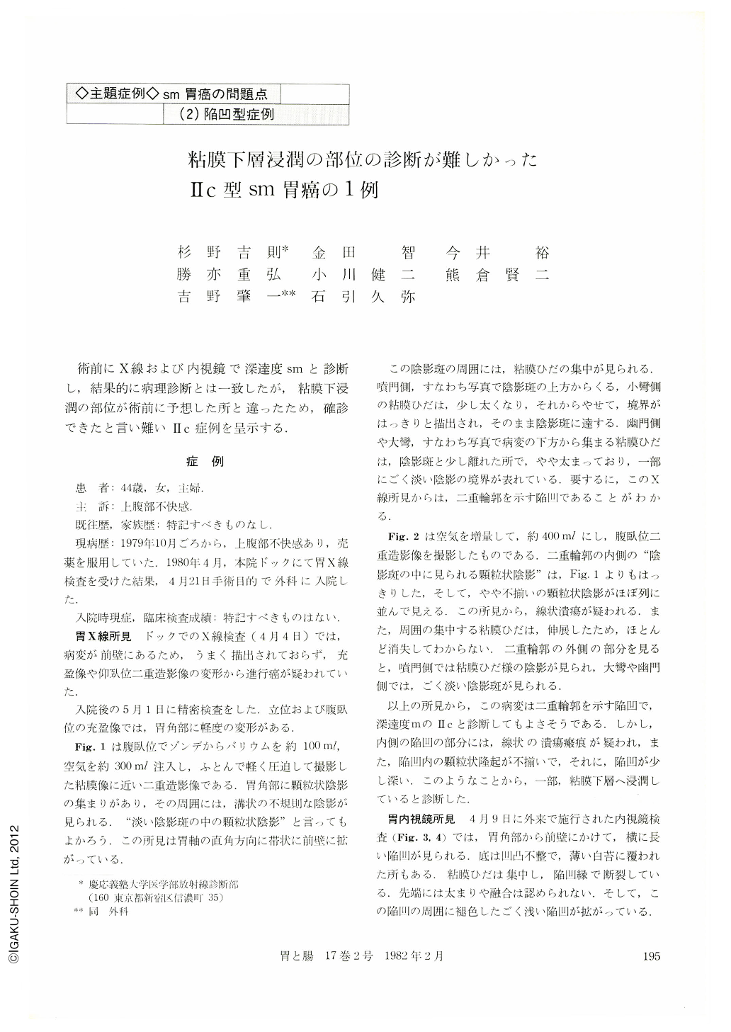
術前にX線および内視鏡で深達度smと診断し,結果的に病理診断とは一致したが,粘膜下浸潤の部位が術前に予想した所と違ったため,確診できたと言い難いⅡc症例を呈示する.
症例
患 者:44歳,女,主婦.
主 訴:上腹部不快感
既往歴,家族歴:特記すべきものなし.
現病歴:1979年10月ごろから,上腹部不快感あり,売薬を服用していた.1980年4月,本院ドックにて胃X線検査を受けた結果,4月21日手術目的で外科に入院した.
The patient, a 44-year-old woman, complained of epigastric discomfort since October 1979. So, she visited Keio Univ. Hospital. The routine upper GI siries on April 4, 1980, revealed an abnormality suggesting gastric cancer. The minute x-ray examination performed on May 1, revealed a wide depressed lesion in the anterior wall of the lower gastric body, which was shallow but double-contoured and multiple granules were recognized within the inner deeper depression associated with a linear ulcer scar.
Endoscopic examination on April 9 showed same findings as those of x-ray. By radiological and endoscopic examination, we diagnosed the lesion as Ⅱc with submucosal involvement. Submucosal invasion of cancer cells was estimated to be accompanied with the linear ulcer scar. Surgical operation was carried out on May, 8, 1980.
The pathological diagnosis of the resected specimen was undifferentiated adenocarcinoma. The size of cancerous lesion was 7.0×6.5 cm. Cancer cells were almost limited in the mucosal layer. Cancerous invasion 0.3 cm in diameter was recognized in the submucosal layer apart from the linear ulcer scar.
The area of submucosal involvement was too small to be recognized macroscopically, so it might be impossible to estimate it radiologically and endoscopically. Meanwhile, by retrospective evaluation of the preoperative findings, there were no definite signs for submucosal involvement.
Copyright © 1982, Igaku-Shoin Ltd. All rights reserved.