Japanese
English
- 有料閲覧
PPV購入案内
1,320円 (1,200円+税10%) こちらは、239ページから243ページの文献です。購入には医書.jp本会員登録が必要です。
会員登録完了後に改めて上記「購入サイトへ」を選択してください。
または「購入サイトへ」選択後に「購入ログイン」、「新規会員登録」の順に進んでください。
会員登録について詳しくはをご確認ください。
- Abstract 文献概要
- 1ページ目 Look Inside
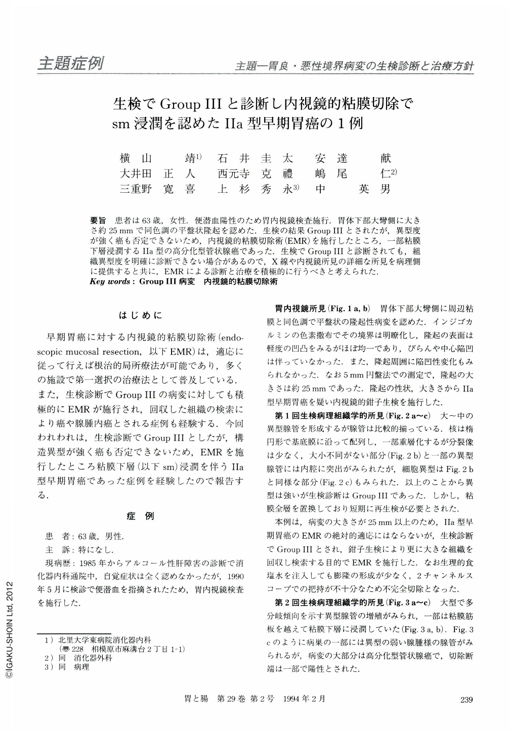
要旨 患者は63歳,女性.便潜血陽性のため胃内視鏡検査施行.胃体下部大彎側に大きさ約25mmで同色調の平盤状隆起を認めた.生検の結果GroupⅢとされたが,異型度が強く癌も否定できないため,内視鏡的粘膜切除術(EMR)を施行したところ,一部粘膜下層浸潤するⅡa型の高分化型管状腺癌であった.生検でGroupⅢと診断されても,組織異型度を明確に診断できない場合があるので,X線や内視鏡所見の詳細な所見を病理側に提供すると共に,EMRによる診断と治療を積極的に行うべきと考えられた.
The patient was a 63-year-old female. Positive fecal occult blood lead to the endoscopic examination which disclosed a flat elevated lesion, about 25 mm in size, on the greater curvature in the lower gastric body. Histological examination of the biopsy specimen showed group Ⅲ. However, cellular atypicality was rather prominent and malignant lesion could not be ruled out. Endoscopic mucosal resection (EMR) was performed for the purpose of both more detailed investigation and treatment. Examination of the endoscopically resected specimen showed that a portion of the tumor invaded the submucosal layer, and the lesion was finally diagnosed as well differentiated tubular adenocarcinoma (type Ⅱa). Even when a gastric lesion is diagnosed as a group Ⅲ disease by conventional biopsy, the presence of cellular atypicality may, nevertheless, suggest a malignant disease. When such lesions are encountered, detailed data obtained from all previous diagnostic studies, such as those from forceps biopsies (several specimens obtained from the same lesion), gastric radiographic and endoscopic examinations (including assessment of the depth of invasion), should be offered to the pathologist. Instead of following up group Ⅲ lesion aimlessly, EMR should be considered at once for the diagnostic and therapeutic purposes.
Copyright © 1994, Igaku-Shoin Ltd. All rights reserved.