Japanese
English
- 有料閲覧
PPV購入案内
1,320円 (1,200円+税10%) こちらは、1557ページから1562ページの文献です。購入には医書.jp本会員登録が必要です。
会員登録完了後に改めて上記「購入サイトへ」を選択してください。
または「購入サイトへ」選択後に「購入ログイン」、「新規会員登録」の順に進んでください。
会員登録について詳しくはをご確認ください。
- Abstract 文献概要
- 1ページ目 Look Inside
- サイト内被引用 Cited by
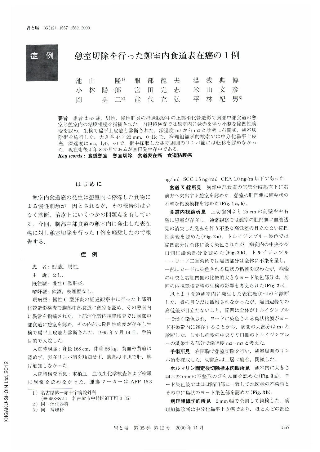
要旨 患者は62歳,男性.慢性肝炎の経過観察中の上部消化管造影で胸部中部食道の憩室と憩室内の粘膜粗糙を指摘された.内視鏡検査では憩室内に発赤を伴う不整な陥凹性病変を認め,生検で扁平上皮癌と診断された.深達度m2からm3と診断し右開胸,憩室切
除術を施行した.大きさ44×22mm,0-Ⅱcで,病理組織学的検索では中分化扁平上皮癌,深達度はm3,ly0,v0で,術中採取した憩室周囲のリンパ節には転移を認めなかった.現在術後4年8か月であるが無再発生存中である.
A 62-year-old male had been followed up for chronic hepatitis, and he was diagnosed by esophagogram as having a middle thoracic esophageal diverticulum with mucosal irregularity. Endoscopically, a reddish and irregular depressed lesion was recognized in the diverticulum. A biopsy specimen showed squamous cell carcinoma. Synthetically, the depth of cancer invasion was estimated as m2 or m3 in the preoperative period, so a diverticulectomy through right thoracotomy was performed. The resected specimen showed a slightly depressed type carcinoma (0-Ⅱc), measuring 44 × 22 mm in size. Histological examination revealed a moderately differentiated squamous cell carcinoma with invasion of m3. Although lymphatic invasion was detected, there was no evidence of venous invasion or lymph node metastasis around the diverticulum. Four years and eight months after surgery, the patient is doing well without any recurrence.
Copyright © 2000, Igaku-Shoin Ltd. All rights reserved.