Japanese
English
- 有料閲覧
PPV購入案内
1,320円 (1,200円+税10%) こちらは、217ページから220ページの文献です。購入には医書.jp本会員登録が必要です。
会員登録完了後に改めて上記「購入サイトへ」を選択してください。
または「購入サイトへ」選択後に「購入ログイン」、「新規会員登録」の順に進んでください。
会員登録について詳しくはをご確認ください。
- Abstract 文献概要
- 1ページ目 Look Inside
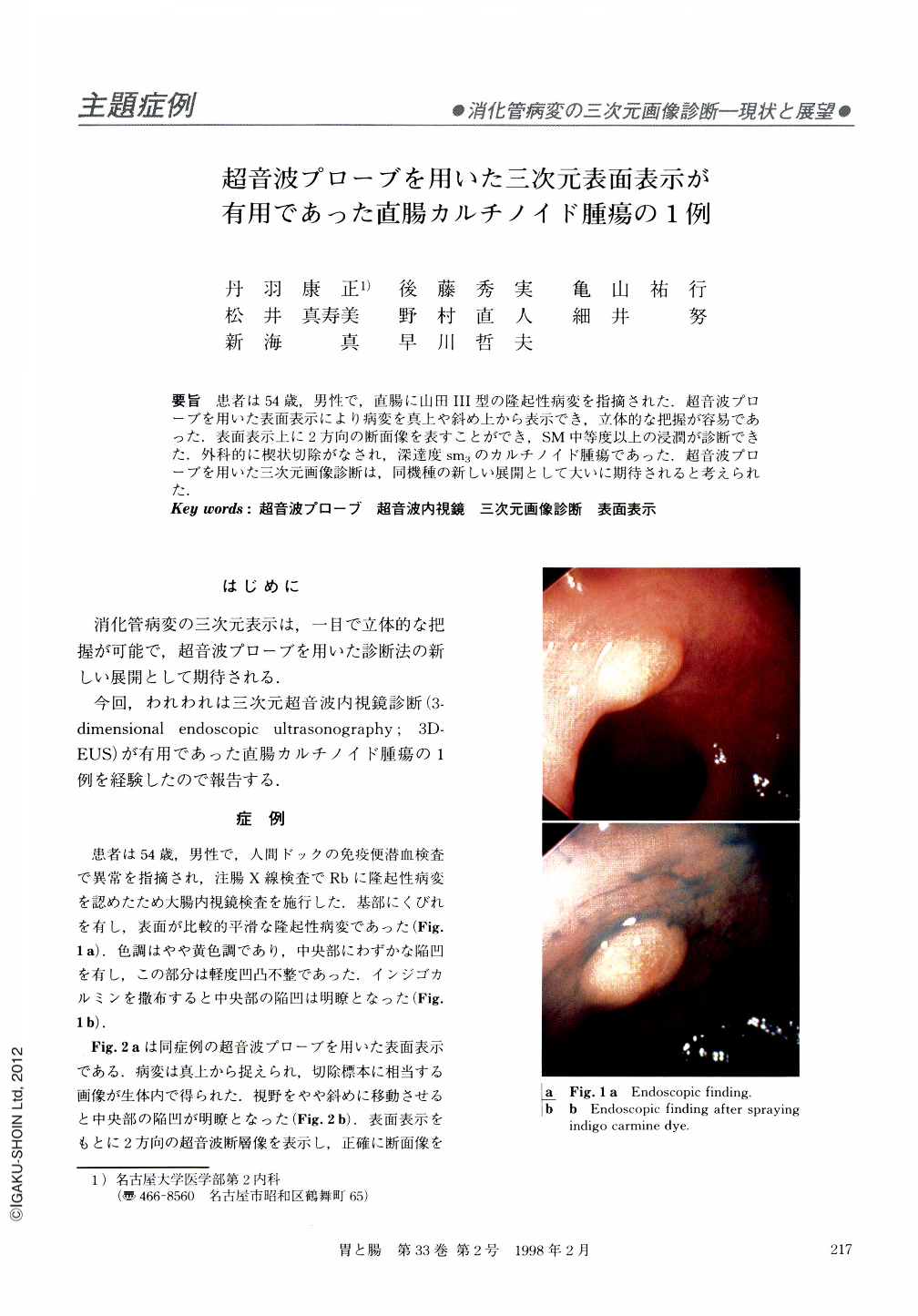
要旨 患者は54歳,男性で,直腸に山田Ⅲ型の隆起性病変を指摘された.超音波プローブを用いた表面表示により病変を真上や斜め上から表示でき,立体的な把握が容易であった.表面表示上に2方向の断面像を表すことができ,SM中等度以上の浸潤が診断できた.外科的に楔状切除がなされ,深達度sm3のカルチノイド腫瘍であった.超音波プローブを用いた三次元画像診断は,同機種の新しい展開として大いに期待されると考えられた.
Following a positive fecal occult blood test in a yearly medical check-up, an elevated lesion in the rectum was discovered in a 54-year-old man. Endoscopic view showed a slightly yellow and smooth surface with an irregular area on the top. Histological biopsy specimen showed carcinoid tumor. The patient underwent ultrasound probe examination while under conventional endoscopic view. Our ultrasound system provided both a surface image and a reconstructed cross-sectional image based on the scan made by the ultrasound probe in water. The reconstructed surface image showed the top view and a slantwise top view. And views can't be obtained from usual endoscopic examination and they were consistent with the macroscopic findings after surgery. The acquisition plane and the reconstructed longitudinal plane were displayed together with the surface image. We were able to know the depth of tumor invasion by the ultrasonographic image overlapped on the surface image. The patient underwent a surgical wedge operation and it was shown that the depth of invasion was as far as the deep submucosal layer. Three-dimensional images using an ultrasound probe are clinically useful and their further use is expected to popularize the method among surgeons.
Copyright © 1998, Igaku-Shoin Ltd. All rights reserved.