Japanese
English
- 有料閲覧
PPV購入案内
1,320円 (1,200円+税10%) こちらは、1413ページから1418ページの文献です。購入には医書.jp本会員登録が必要です。
会員登録完了後に改めて上記「購入サイトへ」を選択してください。
または「購入サイトへ」選択後に「購入ログイン」、「新規会員登録」の順に進んでください。
会員登録について詳しくはをご確認ください。
- Abstract 文献概要
- 1ページ目 Look Inside
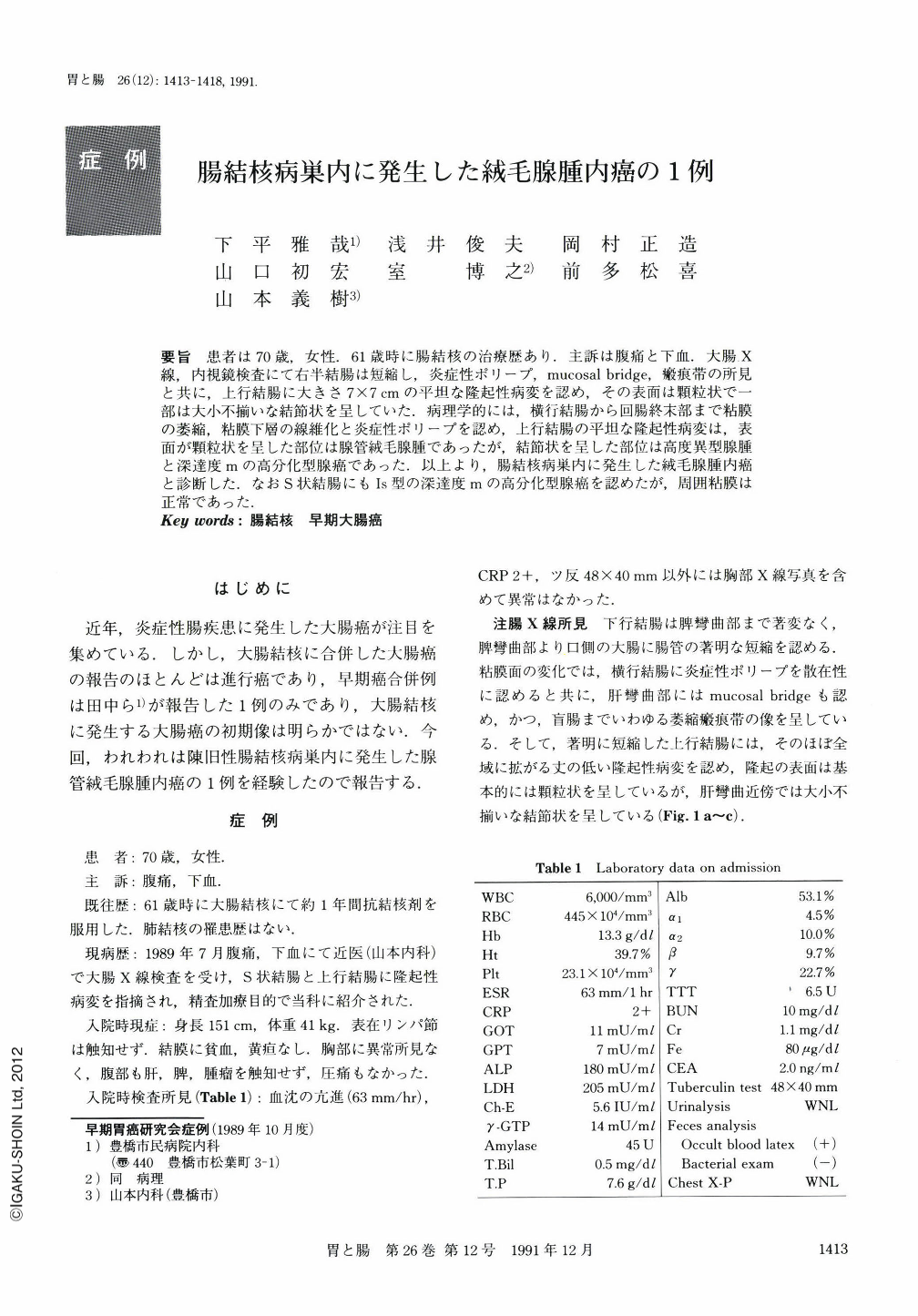
要旨 患者は70歳,女性.61歳時に腸結核の治療歴あり.主訴は腹痛と下血.大腸X線,内視鏡検査にて右半結腸は短縮し,炎症性ポリープ,mucosal bridge,瘢痕帯の所見と共に,上行結腸に大きさ7×7cmの平坦な隆起性病変を認め,その表面は顆粒状で一部は大小不揃いな結節状を呈していた.病理学的には,横行結腸から回腸終末部まで粘膜の萎縮,粘膜下層の線維化と炎症性ポリープを認め,上行結腸の平坦な隆起性病変は,表面が顆粒状を呈した部位は腺管絨毛腺腫であったが,結節状を呈した部位は高度異型腺腫と深達度mの高分化型腺癌であった.以上より,腸結核病巣内に発生した絨毛腺腫内癌と診断した.なおS状結腸にもIs型の深達度mの高分化型腺癌を認めたが,周囲粘膜は正常であった.
A 70-year-old woman with a previous history of intestinal tuberculosis and chief complaints of abdominal pain and melena was pointed out to have abnormalities of the colon on x-ray examination at another hospital and visited our hospital for scrupulous examination. Barium enema and colonoscopic examinations revealed marked shortening and the mucosal atrophy of the right-sided colon. In addition, mucosal tags, inflammatory polyps and absence of haustration were found in the transverse through the ascending colon. A wide and flat elevated lesion with small granular surface pattern was found in the shortened ascending colon. The lesion showed nodular pattern at the hepatic flexure. Another Is type polypoid lesion was detected at the sigmoid colon (Figs. 1 and 2). Right hemicolectomy and partial sigmoidectomy were performed. We diagnosed this lesion at this time as carcinoma in tubulovillous adenoma accompanied with healed colonic tuberculosis.
Macroscopic examination of the resected specimen revealed a flat granular elevated lesion measuring 7×7 cm in the markedly shortened ascending colon and Is type lesion in the sigmoid colon. The mucosa arround the flat granular lesion was atrophic and there was no haustration between the terminal ileum and the transverse colon (Fig. 4).
Histological diagnosis of the flat granular lesion in the ascending colon was well differentiated adenocarcinoma in tubulovillous adenoma limited to the mucosa. The findings of the surrounding mucosa in the transverse colon through the terminal ileum were consistent with healed intestinal tuberculosis. The lesion in the sigmoid colon was also well differentiated adenocarcinoma limited to the mucosa (Figs. 5 and 6).
Based on these findings, this case was considered to be early cancer accompanied with healed colonic tuberculosis.
Copyright © 1991, Igaku-Shoin Ltd. All rights reserved.