Japanese
English
- 有料閲覧
PPV購入案内
1,320円 (1,200円+税10%) こちらは、511ページから514ページの文献です。購入には医書.jp本会員登録が必要です。
会員登録完了後に改めて上記「購入サイトへ」を選択してください。
または「購入サイトへ」選択後に「購入ログイン」、「新規会員登録」の順に進んでください。
会員登録について詳しくはをご確認ください。
- Abstract 文献概要
- 1ページ目 Look Inside
- 参考文献 Reference
- サイト内被引用 Cited by
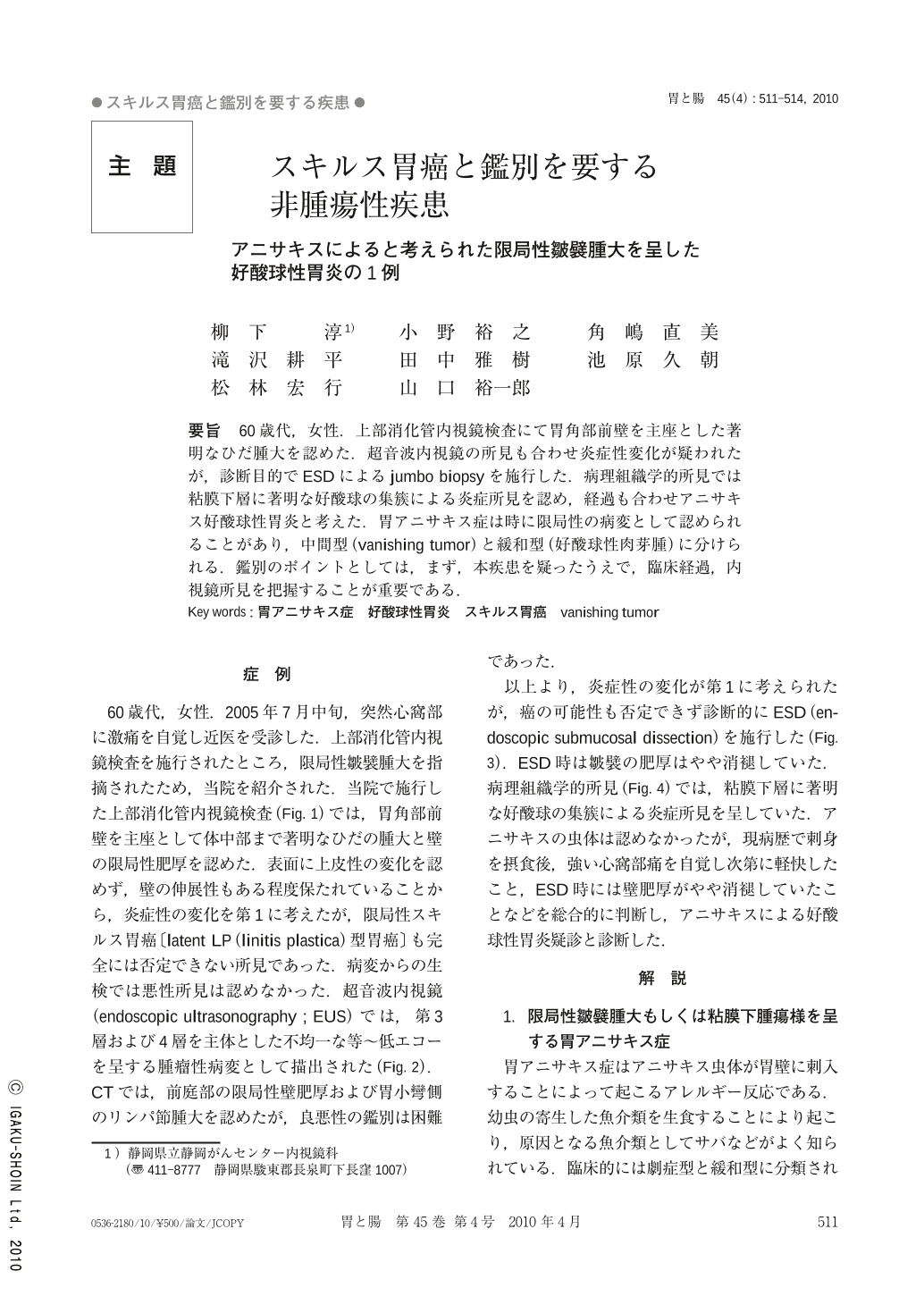
要旨 60歳代,女性.上部消化管内視鏡検査にて胃角部前壁を主座とした著明なひだ腫大を認めた.超音波内視鏡の所見も合わせ炎症性変化が疑われたが,診断目的でESDによるjumbo biopsyを施行した.病理組織学的所見では粘膜下層に著明な好酸球の集簇による炎症所見を認め,経過も合わせアニサキス好酸球性胃炎と考えた.胃アニサキス症は時に限局性の病変として認められることがあり,中間型(vanishing tumor)と緩和型(好酸球性肉芽腫)に分けられる.鑑別のポイントとしては,まず,本疾患を疑ったうえで,臨床経過,内視鏡所見を把握することが重要である.
A woman in her sixties underwent gastroduodenoscopy which revealed limited swelling of the gastric fold of the anterior wall. In addition to the findings of endoscopic ultrasound, the swelling of the gastric fold was considered to be due to inflammation. We performed ESD for the purpose of ensuring that cancer wasn’t present and for making a final diagnosis. Histopathological examination of the ESD specimen revealed inflammation in the submucosal layer by noticeable aggregation of eosinophils. We considered this case as eosinophilic gastritis caused by anisakis. Swelling of the gastric fold or submucosal-tumor-like elevation is infrequent as the endoscopic finding of Gastric anisakiasis. These findings are divided into two types ; vanishing tumor and eosinophic granuloma. For differential diagnosis, first of all, the physician needs to suspect anisakiasis, and then try to find clinical and endoscopic characteristics of anisakiasis.
Copyright © 2010, Igaku-Shoin Ltd. All rights reserved.