Japanese
English
- 有料閲覧
PPV購入案内
1,320円 (1,200円+税10%) こちらは、349ページから355ページの文献です。購入には医書.jp本会員登録が必要です。
会員登録完了後に改めて上記「購入サイトへ」を選択してください。
または「購入サイトへ」選択後に「購入ログイン」、「新規会員登録」の順に進んでください。
会員登録について詳しくはをご確認ください。
- Abstract 文献概要
- 1ページ目 Look Inside
- 参考文献 Reference
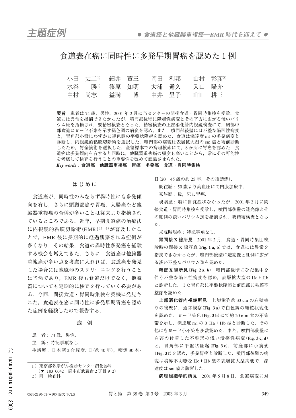
患者は74歳,男性.2001年2月に当センターの間接食道・胃同時集検を受診.食道には異常を指摘できなかったが,噴門部後壁に隆起性病変とその下方に広がる淡いバリウム斑を指摘され,要精密検査となった.精密検査の上部消化管内視鏡検査にて,胸部中部食道にヨード不染を示す褪色調の病変を認め,また,噴門部後壁には不整な陥凹性病変と,胃角部小彎にわずかに褪色調の平盤状隆起を認めた.食道は深達度m1の多発病変と診断し,内視鏡的粘膜切除術を選択した.噴門部の病変は表層拡大型のsm癌と術前診断したため,胃全摘術を選択した.全割標本での病理検索にて,8か所に胃癌を認めた.食道癌は多発傾向を有すると同時に,他臓器重複癌の頻度も高いことから,常にその可能性を考慮して検査を行うことの重要性を改めて認識させられた.
A 74-year-old man underwent gastric mass screening in February, 2001 and a faint barium shadow was detected below an elevated lesion on the posterior wall of the cardiac part, so a detailed examination was made. Endoscopy showed that there were several lesions. One was a pale, iodine-unstained lesion at the middle thoracic esophagus, and another was an irregularly-shaped depression on the posterior wall of the cardiac part. Another was a flat-elevated and slightly pale lesion on the lesser curvature of the angular part. We diagnosed the depth of invasion of the lesions at the esophagus as Tis (M 1), so endoscopic mucosal resection (EMR) was performed. We diagnosed the depth of invasion of the superficial spreading tumor on the posterior wall of the cardiac part as T 1 (SM), so total gastrectomy was performed. In the histological examination of the resected specimen, eight early gastric cancers were revealed. When making examinations, it is very important to make allowances for the possibility of not only multiple esophageal cancers but also double cancers of other organs.
Copyright © 2003, Igaku-Shoin Ltd. All rights reserved.