Japanese
English
- 有料閲覧
PPV購入案内
1,320円 (1,200円+税10%) こちらは、200ページから213ページの文献です。購入には医書.jp本会員登録が必要です。
会員登録完了後に改めて上記「購入サイトへ」を選択してください。
または「購入サイトへ」選択後に「購入ログイン」、「新規会員登録」の順に進んでください。
会員登録について詳しくはをご確認ください。
- Abstract 文献概要
- 1ページ目 Look Inside
- 参考文献 Reference
- サイト内被引用 Cited by
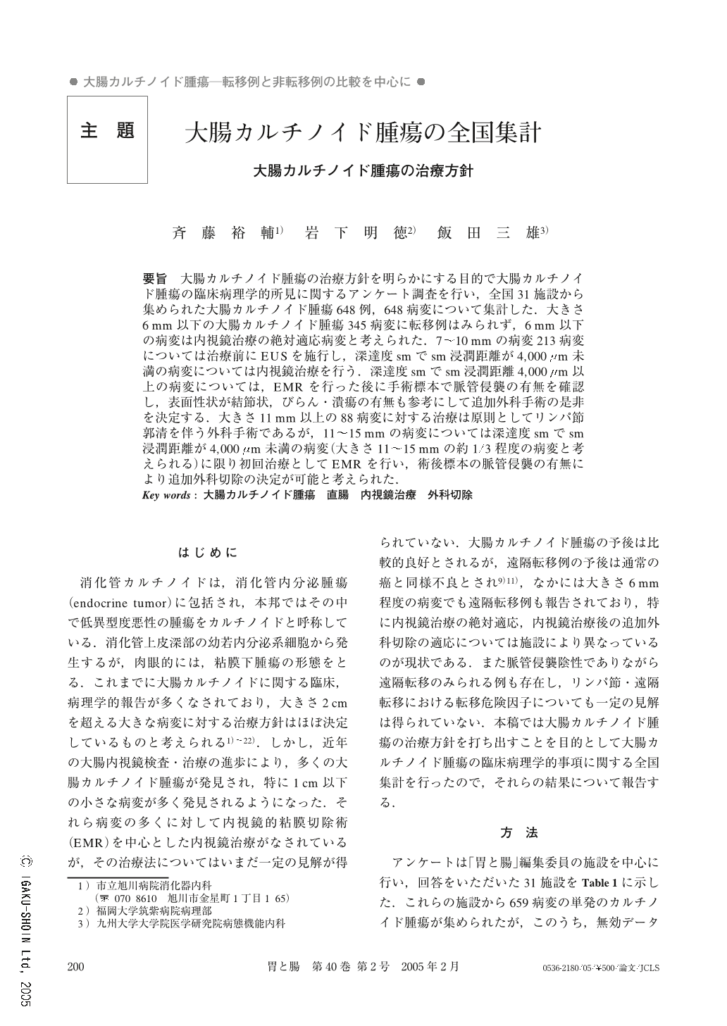
要旨 大腸カルチノイド腫瘍の治療方針を明らかにする目的で大腸カルチノイド腫瘍の臨床病理学的所見に関するアンケート調査を行い,全国31施設から集められた大腸カルチノイド腫瘍648例,648病変について集計した.大きさ6mm以下の大腸カルチノイド腫瘍345病変に転移例はみられず,6mm以下の病変は内視鏡治療の絶対適応病変と考えられた.7~10mmの病変213病変については治療前にEUSを施行し,深達度smでsm浸潤距離が4,000μm未満の病変については内視鏡治療を行う.深達度smでsm浸潤距離4,000μm以上の病変については,EMRを行った後に手術標本で脈管侵襲の有無を確認し,表面性状が結節状,びらん・潰瘍の有無も参考にして追加外科手術の是非を決定する.大きさ11mm以上の88病変に対する治療は原則としてリンパ節郭清を伴う外科手術であるが,11~15mmの病変については深達度smでsm浸潤距離が4,000μm未満の病変(大きさ11~15mmの約1/3程度の病変と考えられる)に限り初回治療としてEMRを行い,術後標本の脈管侵襲の有無により追加外科切除の決定が可能と考えられた.
In order to elucidate the therapeutic strategy for colorectal carcinoid tumors, 648 lesions of 648 cases colorectal carcinoid tumors were collected and analyzed through a quentionnaire survey of 31 institutions in Japan. Carcinoid tumors 6 mm or less in size can be treated by endoscopic therapy alone (endoscopic mucosal resection : EMR) because they have neither lymphnode nor distant metastasis. For the lesions 7~10 mm in size, EUS is recommended and submucosal invasion depth should be measured prior to determination of therapy. Lesions with less than 4,000μm submucosal invasion depth can be treated by EMR alone. However, the lesions with 4,000μm or more submucosal invasion depth can be initially treated by EMR, but additional surgery should be considered for the lesions with lymphatic or venous permeation in post EMR specimens. Additional surgery should be also considered for nodular shaped lesions or lesions with surface ulcers. For the lesions with 11 mm or more in diameter, initial surgical operation with lymphadenectomy should be selected. If the size of the lesion is 11~15 mm in size and if it is diagnosed as a submucosal lesion with a submucosal invasion depth less than 4,000μm (about 1/3 of lesions are 11~15 mm in size), it can be initially resected by EMR and additional surgery should be considered if lymphatic or venous permeation is detected in the post EMR specimens.
Copyright © 2005, Igaku-Shoin Ltd. All rights reserved.