Japanese
English
- 有料閲覧
PPV購入案内
1,320円 (1,200円+税10%) こちらは、1239ページから1254ページの文献です。購入には医書.jp本会員登録が必要です。
会員登録完了後に改めて上記「購入サイトへ」を選択してください。
または「購入サイトへ」選択後に「購入ログイン」、「新規会員登録」の順に進んでください。
会員登録について詳しくはをご確認ください。
- Abstract 文献概要
- 1ページ目 Look Inside
- 参考文献 Reference
- サイト内被引用 Cited by
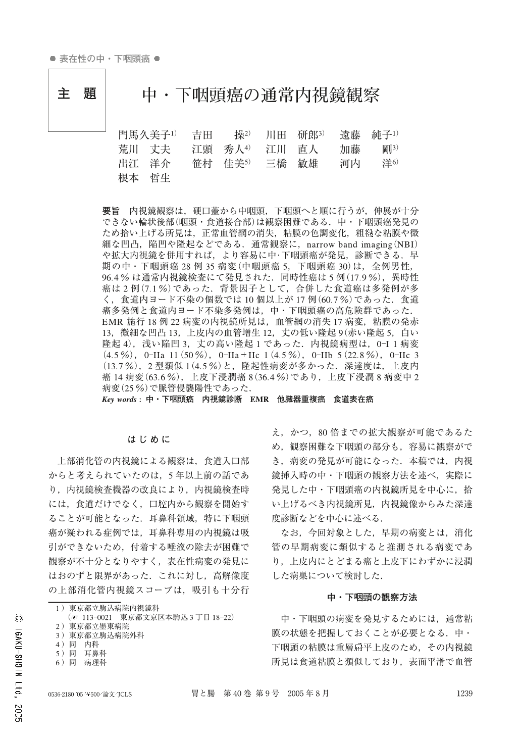
要旨 内視鏡観察は,硬口蓋から中咽頭,下咽頭へと順に行うが,伸展が十分できない輪状後部(咽頭・食道接合部)は観察困難である.中・下咽頭癌発見のため拾い上げる所見は,正常血管網の消失,粘膜の色調変化,粗そうな粘膜や微細な凹凸,陥凹や隆起などである.通常観察に,narrow band imaging(NBI)や拡大内視鏡を併用すれば,より容易に中・下咽頭癌が発見,診断できる.早期の中・下咽頭癌28例35病変(中咽頭癌5,下咽頭癌30)は,全例男性,96.4%は通常内視鏡検査にて発見された.同時性癌は5例(17.9%),異時性癌は2例(7.1%)であった.背景因子として,合併した食道癌は多発例が多く,食道内ヨード不染の個数では10個以上が17例(60.7%)であった.食道癌多発例と食道内ヨード不染多発例は,中・下咽頭癌の高危険群であった.EMR施行18例22病変の内視鏡所見は,血管網の消失17病変,粘膜の発赤13,微細な凹凸13,上皮内の血管増生12,丈の低い隆起9(赤い隆起5,白い隆起4),浅い陥凹3,丈の高い隆起1であった.内視鏡病型は,0-I 1病変(4.5%),0-IIa1 1(50%),0-IIa+IIc 1(4.5%),0-IIb 5(22.8%),0-IIc 3(13.7%),2型類似1(4.5%)と,隆起性病変が多かった.深達度は,上皮内癌14病変(63.6%),上皮下浸潤癌8(36.4%)であり,上皮下浸潤8病変中2病変(25%)で脈管侵襲陽性であった.
In surveillance of carcinoma of the mesopharynx and the hypopharynx, keen observation is recommended as the scope is introduced and also as it is withdrawn. Endoscopic observation should be started as soon as the scope is introduced into the mouth. There are some difficulties in observation of mucosal lesions in the post-cricoid area. The narrow band imaging (NBI) system will make surveillance easy, because it allows us to differentiate a cancer as a mucosal lesion with color different from normal pharyngeal mucosa. Magnifying endoscopy is also useful for differentiation of mucosal lesions. Thirty-five early-stage cancer lesions among 28 patients were identified at the mesopharynx (5) and the hypopharynx (30). Most of them (96.4% of all lesions) were found by upper GI endoscopy. Five patients (17.9%) had synchronous multiple cancer lesions and two cases had metachronous multiple cancer lesions. Cancer lesions at the mesopharynx and the hypopharynx were frequent among patients with multiple esophageal cancers and whose esophageal mucosa had many iodine unstained areas. Ten patients had undergone irradiation until 2001 and another 18 patients had undergone endoscopic mucosal resection (EMR) after 2002. Endoscopic findings of lesions treated by EMR were interrupted mucosal vessels (17 lesions), reddening of the mucosa (13), fine granular changes (13), proliferated mucosal vessels (12), slight elevation (9 : 5 with reddening and 4 with white elevation), protruding lesion 1 and slight depression (3). Sizes of cancer lesions were from 6 mm to 40 mm. Overall, elevated lesions were in the majority, such as type 0-I 1 lesion (4.5%), 0-IIa 11 (50%), 0-IIa+IIc 1 (4.5%), 0-IIb 5 (22.8%), 0-IIc 3 (13.7%) and type 2 1 (4.5%). Fourteen lesions (63.6%) remained within the epithelium and 8 lesions (36.4%) showed invasion into the subepithelial tissue. Microvascular permeation was identified in two cases (25%) of eight which had invasion into the subepithelial layer.
Copyright © 2005, Igaku-Shoin Ltd. All rights reserved.