Japanese
English
- 有料閲覧
PPV購入案内
1,320円 (1,200円+税10%) こちらは、466ページから473ページの文献です。購入には医書.jp本会員登録が必要です。
会員登録完了後に改めて上記「購入サイトへ」を選択してください。
または「購入サイトへ」選択後に「購入ログイン」、「新規会員登録」の順に進んでください。
会員登録について詳しくはをご確認ください。
- Abstract 文献概要
- 1ページ目 Look Inside
- 参考文献 Reference
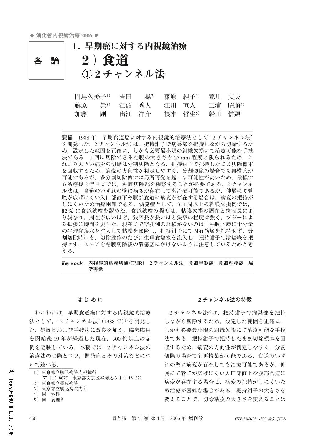
要旨 1988年,早期食道癌に対する内視鏡的治療法として“2チャンネル法”を開発した.2チャンネル法は,把持鉗子で病巣部を把持しながら切除するため,設定した範囲を正確に,しかも必要最小限の組織欠損にて治療可能な手技法である.1回に切除できる粘膜の大きさが25mm程度と限られるため,これより大きい病変の切除は分割切除となる.把持鉗子で把持したまま切除標本を回収するため,病変の方向性が判定しやすく,分割切除の場合でも再構築が可能であるが,多分割切除例では局所再発を起こす可能性が高いため,最低でも治療後2年目までは,粘膜切除部を観察することが必要である.2チャンネル法は,食道のいずれの壁に病変が存在しても治療可能であるが,伸展にて管腔が広げにくい入口部直下や腹部食道に病変が存在する場合は,病変の把持がしにくいため治療困難である.偶発症として,3/4周以上の粘膜欠損例では,82%に食道狭窄を認めた.食道狭窄の程度は,粘膜欠損の周在と狭窄長により異なり,周在が広いほど,狭窄長が長いほど狭窄の程度は強く,ブジーによる拡張に時間を要した.現在まで穿孔例の経験がないのは,粘膜下層に十分量の生理食塩水を注入して粘膜を膨隆し,把持鉗子にて固有筋層を把持せず,分割切除時にも,切除操作のたびに生理食塩水を注入し,把持鉗子で潰瘍底を把持せず,スネアを粘膜切除後の潰瘍底にかけないように注意しているためと考える.
We reported the first paper on endoscopic mucosal resection (EMR) by “the two-channel technique” as a treatment of mucosal cancer of the esophagus. An EMR using the two-channel technique allowed us to remove a mucosal lesion with the lining submucosa as we intended and to keep the mucosal defect minimum. Because the maximal dimension of resected mucosa is 25 mm, any mucosal lesion larger than 25 mm must be removed by repeated resections until the lesion is removed completely. This technique also allowed us to reconfirm the mutual relationship between the resected specimens. To do this, we kept every specimen as soon as it had been collected by the forceps. It was recommended that any patient who underwent EMR with piecemeal resection should be kept under endoscopic surveillance against local recurrence at least for two years after the treatment, because the incidence of local recurrence after EMR is significantly high among patients treated by piecemeal resection. The two-channel technique can be applied in any mucosal resection at any part of the esophagus, except mucosal lesions at the pharyngoesophageal junction or at the distal esophagus close to the esophagogastric junction where procedures for EMR are difficult because of the narrow lumen. As a complication immediately due to EMR, esophageal wall perforation is most severe. The methods of prevention of esophageal wall perforation were as follows ; to inject a sufficient amount of normal saline into the submucosa and to elevate the mucosal lesion sufficiently immediately before every resection and to avoid grasping the proper muscle layer by the forceps especially in cases involving piecemeal resection. These measures enabled us to avoid the esophageal wall perforation by EMR. Esophageal stenosis was one of the important late complications. It was frequent (82%) among patients whose mucosal defect occupied over 3/4 of the circumference of the esophagus. The grade of stenosis increased as the mucosal defect increased in size either in circumference or in axial length. Dilatation during treatment took a longer period when there was severe stenosis.
Copyright © 2006, Igaku-Shoin Ltd. All rights reserved.