Japanese
English
- 有料閲覧
- Abstract 文献概要
- 1ページ目 Look Inside
- 参考文献 Reference
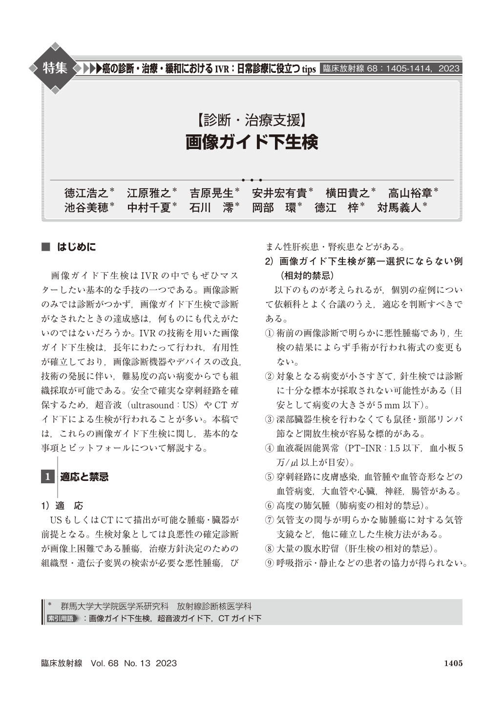
画像ガイド下生検はIVRの中でもぜひマスターしたい基本的な手技の一つである。画像診断のみでは診断がつかず,画像ガイド下生検で診断がなされたときの達成感は,何ものにも代えがたいのではないだろうか。IVRの技術を用いた画像ガイド下生検は,長年にわたって行われ,有用性が確立しており,画像診断機器やデバイスの改良,技術の発展に伴い,難易度の高い病変からでも組織採取が可能である。安全で確実な穿刺経路を確保するため,超音波(ultrasound:US)やCTガイド下による生検が行われることが多い。本稿では,これらの画像ガイド下生検に関し,基本的な事項とピットフォールについて解説する。
With the improvement of diagnostic imaging devices and the development of technology, various tissue samples can be collected by image-guided biopsy. In recent years, cancer treatment has been progressing toward “personalized medicine,” in which treatment is based on cancer gene information, in addition to surgery and radiation therapy. Appropriate specimen collection is essential not only for cancer diagnosis but also for cancer gene analysis for drug selection, requiring minimally invasive, safe, and highly accurate image-guided biopsy before treatment. Image-guided biopsy is particularly important to ensure a safe and reliable puncture route. This article describes the basics and pitfalls of image-guided biopsy.

Copyright © 2023, KANEHARA SHUPPAN Co.LTD. All rights reserved.


