Japanese
English
- 有料閲覧
- Abstract 文献概要
- 1ページ目 Look Inside
- 参考文献 Reference
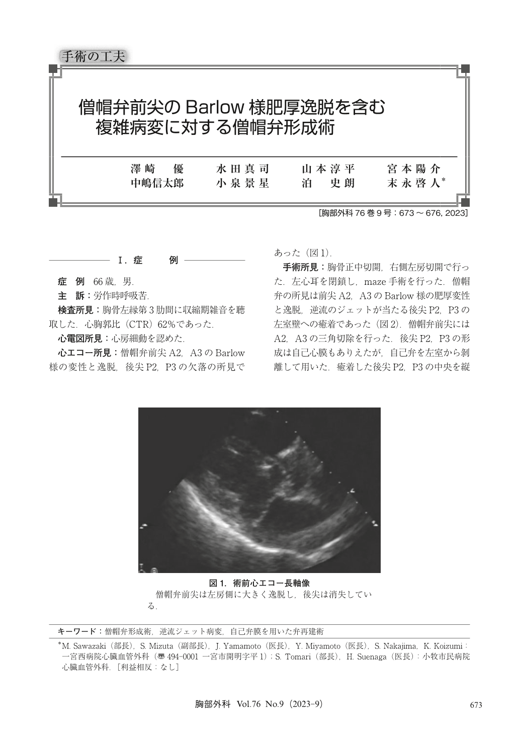
1)前尖の肥厚変性病変による僧帽弁閉鎖不全症の逆流ジェットが当たる後尖P2,P3が左室に癒着した症例の形成術式を報告した.2)後尖弁膜の癒着をはがし逆T字切開し,切開した部分の弁輪を縫縮し,砂時計切除3)の逆で,弁尖の高さを増し膨らみをもたせるという術式である.自己弁組織は耐久性に優れ,使える場合には優先して使用したい.
A 66 year-old male was admitted to our clinic suffering from dyspnea on effort. Cardio thoracic ratio (CTR) was 62%. Electrocardiogram showed atrial fibrillation. Echocardiogram showed severe mitral regurgitation (MR), Barlow like billowing and thickened A2 and A3, and loss of P2 and P3. Operation was performed through median sternotomy and right sided left atrial incision. Left atrial appendage was closed with running suture. Maze operation was done. Triangular resection of A2 and A3 was done. P2 and P3 were adhered to the left ventricular wall. First we cut the adhered posterior leaflet in a shape of inverted T. And the adhered leaflet was dissected from the left ventricle by the scissors. The detached annulus was mattress-sutured with a pledgetted suture. The leaflets were sutured together, then a new posterior leaflet was remade using mitral valve leaflet tissue and the shape became higher and round. Post operatively, MR was none, and posterior leaflet functioned well. Sinus rhythm was recovered. Eleven years later, no MR and sinus rhythm were shown.

© Nankodo Co., Ltd., 2023


