Japanese
English
臨床経験
増大経過を5年間CT所見で追跡しえた囊胞状肺癌
Growth Process of Cystic Lung Cancer Followed with Computed Tomography Findings Over 5 Years
笠井 由隆
1
,
伊藤 公一
1
,
桝屋 大輝
1
,
久米 佐知枝
1
,
井上 明香
1
,
鈴木 雄二郎
1
Yoshitaka Kasai
1
,
Kouichi Ito
1
,
Daiki Masuya
1
,
Sachie Kume
1
,
Sayaka Inoue
1
,
Yuichiro Suzuki
1
1神鋼記念病院呼吸器センター
1Department of Respiratory Center, Shinko Memorial Hospital
キーワード:
原発性肺癌
,
薄壁空洞
,
肺囊胞
,
チェックバルブ
Keyword:
primary lung cancer
,
thin-walled cavity
,
pulmonary cyst
,
check valve
pp.336-338
発行日 2018年5月1日
Published Date 2018/5/1
DOI https://doi.org/10.15106/j_kyobu71_336
- 有料閲覧
- Abstract 文献概要
- 1ページ目 Look Inside
- 参考文献 Reference
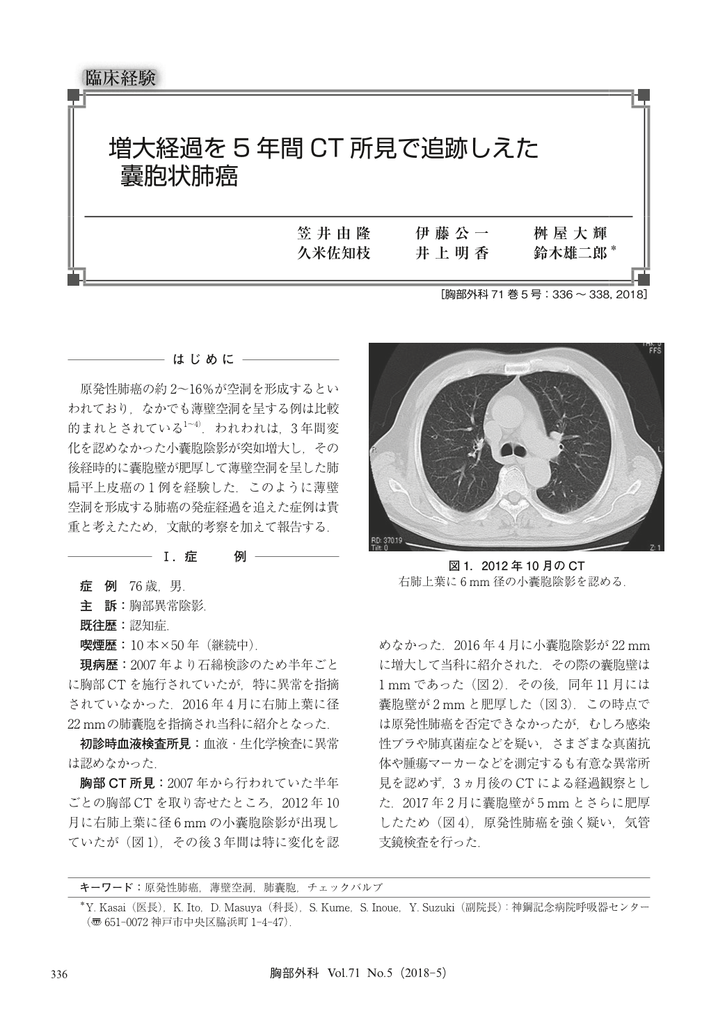
原発性肺癌の約2~16%が空洞を形成するといわれており,なかでも薄壁空洞を呈する例は比較的まれとされている1~4).われわれは,3年間変化を認めなかった小囊胞陰影が突如増大し,その後経時的に囊胞壁が肥厚して薄壁空洞を呈した肺扁平上皮癌の1例を経験した.このように薄壁空洞を形成する肺癌の発症経過を追えた症例は貴重と考えたため,文献的考察を加えて報告する.
An estimated 2~16% of primary lung cancers form cavities with cases that form thin-walled cavities being comparatively rare. We treated a patient with squamous cell carcinoma of the lung with a small cystic shadow that showed no changes for 3 years. The cyst then suddenly grew larger, after which the cyst wall thickened over time and a thin-walled cavity was seen. Here we report this important case showing the development process of lung cancer that formed a thin-walled cavity, together with a discussion of the literature.

© Nankodo Co., Ltd., 2018


