Japanese
English
- 有料閲覧
- Abstract 文献概要
- 1ページ目 Look Inside
- 参考文献 Reference
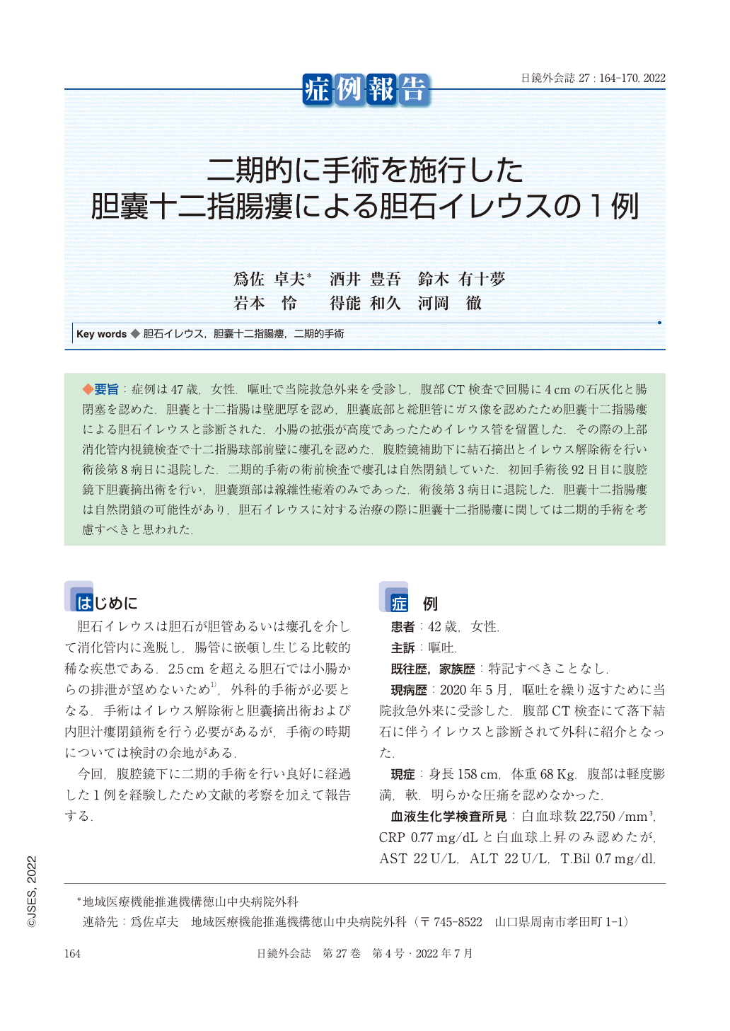
◆要旨:症例は47歳,女性.嘔吐で当院救急外来を受診し,腹部CT検査で回腸に4cmの石灰化と腸閉塞を認めた.胆囊と十二指腸は壁肥厚を認め,胆囊底部と総胆管にガス像を認めたため胆囊十二指腸瘻による胆石イレウスと診断された.小腸の拡張が高度であったためイレウス管を留置した.その際の上部消化管内視鏡検査で十二指腸球部前壁に瘻孔を認めた.腹腔鏡補助下に結石摘出とイレウス解除術を行い術後第8病日に退院した.二期的手術の術前検査で瘻孔は自然閉鎖していた.初回手術後92日目に腹腔鏡下胆囊摘出術を行い,胆囊頸部は線維性癒着のみであった.術後第3病日に退院した.胆囊十二指腸瘻は自然閉鎖の可能性があり,胆石イレウスに対する治療の際に胆囊十二指腸瘻に関しては二期的手術を考慮すべきと思われた.
A 40-year-old female who complained of vomiting was admitted to our hospital. Abdominal CT revealed a 4 cm calcification in the small bowel that caused bowel obstruction. Additional findings were gas in the common bile duct and the gallbladder with the fistula between the gallbladder and the duodenum. The diagnosis of gallstone ileus with the cholecystoduodenal fistula was made. As her general condition was good, we planned the two-stage surgery and put the ileus tube with esophagogastroduodenoscopy (EGD) which showed the cholecystoduodenal fistula at the anterior wall of the duodenal bulb. In the first operation, laparoscopy easily revealed the small bowel obstruction due to the stone. The small intestine was pulled out and the stone was extracted after cutting the small intestine. On the postoperative day 8, the patient was discharged. Before the second operation, EGD showed that the fistula was closed, and CT showed the gallbladder was atrophic and edematous. After 92 days from the first operation, laparoscopic cholecystectomy was performed. No fistula was observed during surgery. The patient was discharged on postoperative day 3. The two-stage surgery should be considered as a treatment for gallstone ileus with cholecystoduodenal fistula because this fistula may be spontaneously.

Copyright © 2022, JAPAN SOCIETY FOR ENDOSCOPIC SURGERY All rights reserved.


