Japanese
English
- 有料閲覧
- Abstract 文献概要
- 1ページ目 Look Inside
- 参考文献 Reference
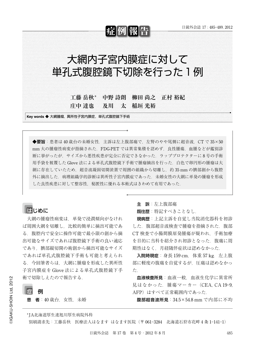
◆要旨:患者は40歳台の未婚女性.主訴は左上腹部痛で,左腎のやや尾側に超音波,CTで35×50mm大の腫瘤性病変が指摘された.FDG-PETでは異常集積を認めず,良性腫瘍,血腫などが鑑別診断に挙がったが,サイズから悪性疾患が完全に否定できなかった.ラッププロテクターに8号の手術用手袋を被覆したGlove法による単孔式腹腔鏡下手術で腫瘤摘出を行った.白色で卵円形の腫瘤は大網に存在していたため,超音波凝固切開装置で周囲の組織から切離し,約35mmの臍部創から腹腔外に摘出した.病理組織学的診断は異所性子宮内膜症であった.未婚女性の大網に単発の腫瘤を形成した良性疾患に対して整容性,秘匿性に優れる本術式はきわめて有用であった.
A woman in her forties had left upper abdominal pain and came to our hospital. Abdominal ultrasonography and CT showed a 35×50mm mass caudad to the left kidney. FDG-PET showed that the mass had no abnormal uptake. Differential diagnosis was benign tumor or hematoma, however, potential malignancy of the mass could not be completely denied because of its size. The mass was resected by single incision laparoscopic surgery using Lap-Protector wrapped with surgical glove. Using laparoscopic coagulating scissors, the mass, which was actually located in the great omentum, was parted from surrounding tissue and taken out of the abdominal space through an approximately 35 mm vertical umbilical incision. Pathological diagnosis of the mass was endometriosis heterotopica. Single incision laparoscopic surgery was very useful for resecting the benign, single mass which existed in the great omentum of the unmarried female patient, and for its cosmesis and concealment.

Copyright © 2012, JAPAN SOCIETY FOR ENDOSCOPIC SURGERY All rights reserved.


