Japanese
English
- 有料閲覧
- Abstract 文献概要
- 1ページ目 Look Inside
Ⅰ.緒言
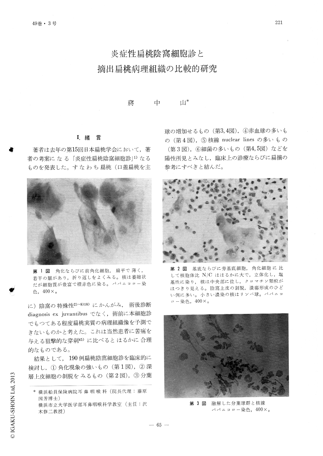
著者は去年の第15回日本扁桃学会において,著者の考案になる「炎症性扁桃陰窩細胞診」1)なるものを発表した。すなわち扁桃(口蓋扁桃を主に)陰窩の特殊性2)〜8)18)にかんがみ,術後診断diagnosis ex juvantibusでなく,術前に本細胞診でもつてある程度扁桃実質の病理組織像を予測できないものかと考えた。これは当然患者に苦痛を与える狙撃的な穿刺45)に比べるとはるかに合理的なものである。
結果として,190例扁桃陰窩細胞診を臨床的に検討し,①角化現象の強いもの(第1図),②深層上皮細胞の剥脱をみるもの(第2図),③分葉球の増加せるもの(第3,4図),④赤血球の多いもの(第4図),⑤核線nuclear linesの多いもの(第3図),⑥細菌の多いもの(第4,5図)などを陽性所見とみなし,臨床上の診療ならびに扁摘の参考にすべきと結んだ。
From my first trial on, the prospection of preoperative tonsillar pathological histology by the simple method of cytology-the previously reported, "Clinical Studies on Inflammatory Tonsillar Lacunar Cytology", (Otologia, Fukuoka, 22:210-224, 1976) I came to the conclusion that those positive findings such as, (1) strong cornification, (2) exfoliation of the deeper epithelial cells, (3) increase of segments, (4) incr. ease of R. B. C., (5) increase of nuclear lines, (6) increase of bacterial count etc. are helpful in the determination of clinical treatment or removal of tonsils.
On the other hand, in contrast to those which were mainly based on clinical findings and studies, I have devised another method, through studies on 80 extirpated tonsils, to prove that those positive tonsillar lacunar cytological findings are in positive correlation with the severeness of the pathological changes.
These tonsils were classified into, IInd type of atrophic, IIIrd type degenerative, IVth type of atrophic and sclerosing phases and, with 2 in termediate types of IInd and IIIrd, and IIIrd and I Vth.
Every case was analysed in detail of their pathological findings and designated with their tonsillar pathological coefficient in accordance to its coefficient table.

Copyright © 1977, Igaku-Shoin Ltd. All rights reserved.


