Japanese
English
- 有料閲覧
- Abstract 文献概要
- 1ページ目 Look Inside
I.はじめに
頭部打撲後,両耳側半盲を呈することは稀ながら文献上散見され,その発生要因として何らかの視交叉部の障害が指摘されているが,その手術所見の報告は稀である.
今回われわれは頭蓋底骨折に伴い両耳側半盲を認め,髄液漏根治術の際に視交叉部周辺を観察し得た1例を経験した.その臨床経過,画像所見,術中所見より両耳側半盲の原因を検討し,発生機序を中心に若干の文献的考察を加え報告する.
We report a case of bitemporal hemianopsia after skullbase fracture.
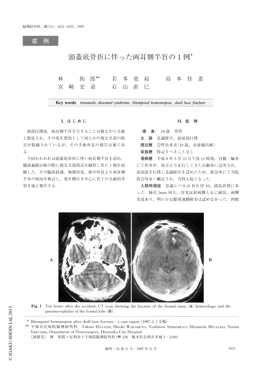
An 18-year-old male presented with frontal head hit due to a traffic accident. Consciousness level on admis-sion was JCS 10. Initial CT scan revealed traumatic SAH and pneumocephalus. We treated him conserva-tively. Ten hours after the accident, consciousness level went down, and CT imaging disclosed bilateral frontal hemorrhage. The three dimensional CT (3D-CT) imag-ing showed two fracture lines from the roof of the ethmoid sinus to the planum sphenoidale. Although his consciousness improved gradually, he complained of rhinorrhea, anosmia and double vision. On pre-operative visual field examination, bitemporal hemi-anopsia was noticed. Repair operation for CSF rhinor-rhea was performed. Operative findings revealed two fracture lines corresponding to the 3D-CT scan. The optic chiasm was compressed by the tuberculum sellae. We could not find obvious tearings or stretchings of the chiasm.
Reports on operative findings of traumatic chiasmal syndrome are rare, and most of the reports presume that bitemporal hemianopsia results from tearing or stretching of the chiasm. We could confirm that, in some cases, bitemporal hemianopsia could result from direct pressure of the tuberculum sellae.

Copyright © 1997, Igaku-Shoin Ltd. All rights reserved.


