Japanese
English
症例
頸椎Aneurysmal Bone Cystの1例
Aneurysmal Bone Cyst of the Sixth Cervical Spine:Case report
岡田 尚巳
1
,
長谷川 光広
1
,
山嶋 哲盛
1
,
山下 純宏
1
,
木谷 隆一
2
Takashi OKADA
1
,
Mitsuhiro HASEGAWA
1
,
Tetsumori YAMASHIMA
1
,
Junkoh YAMASHITA
1
,
Ryuuiti KITANI
2
1金沢大学脳神経外科
2富山労災病院脳神経外科
1Department of Neurosurgery, School of Medicine, Kanazawa University
2Department of Neurosurgery, Toyama Rohsai Hospital
キーワード:
Aneurysmal bone cyst
,
Cervical spine
,
Magnetic resonance imaging
Keyword:
Aneurysmal bone cyst
,
Cervical spine
,
Magnetic resonance imaging
pp.1043-1047
発行日 1993年11月10日
Published Date 1993/11/10
DOI https://doi.org/10.11477/mf.1436900743
- 有料閲覧
- Abstract 文献概要
- 1ページ目 Look Inside
I.緒言
Aneurysmal Bone Cyst(以下ABC)は良性腫瘍性骨疾患に分類され全骨腫瘍の約1%を占める13.長管骨に好発するが,脊椎にも発生し,腰椎,胸椎の順にみられる.頸椎に発生するABCは,これまで本邦では9例の報告がみられるに過ぎない、今回われわれは,第6頸椎に限局してみられたABCの1治験例を経験したので,若干の文献的考察を加えて報告する.
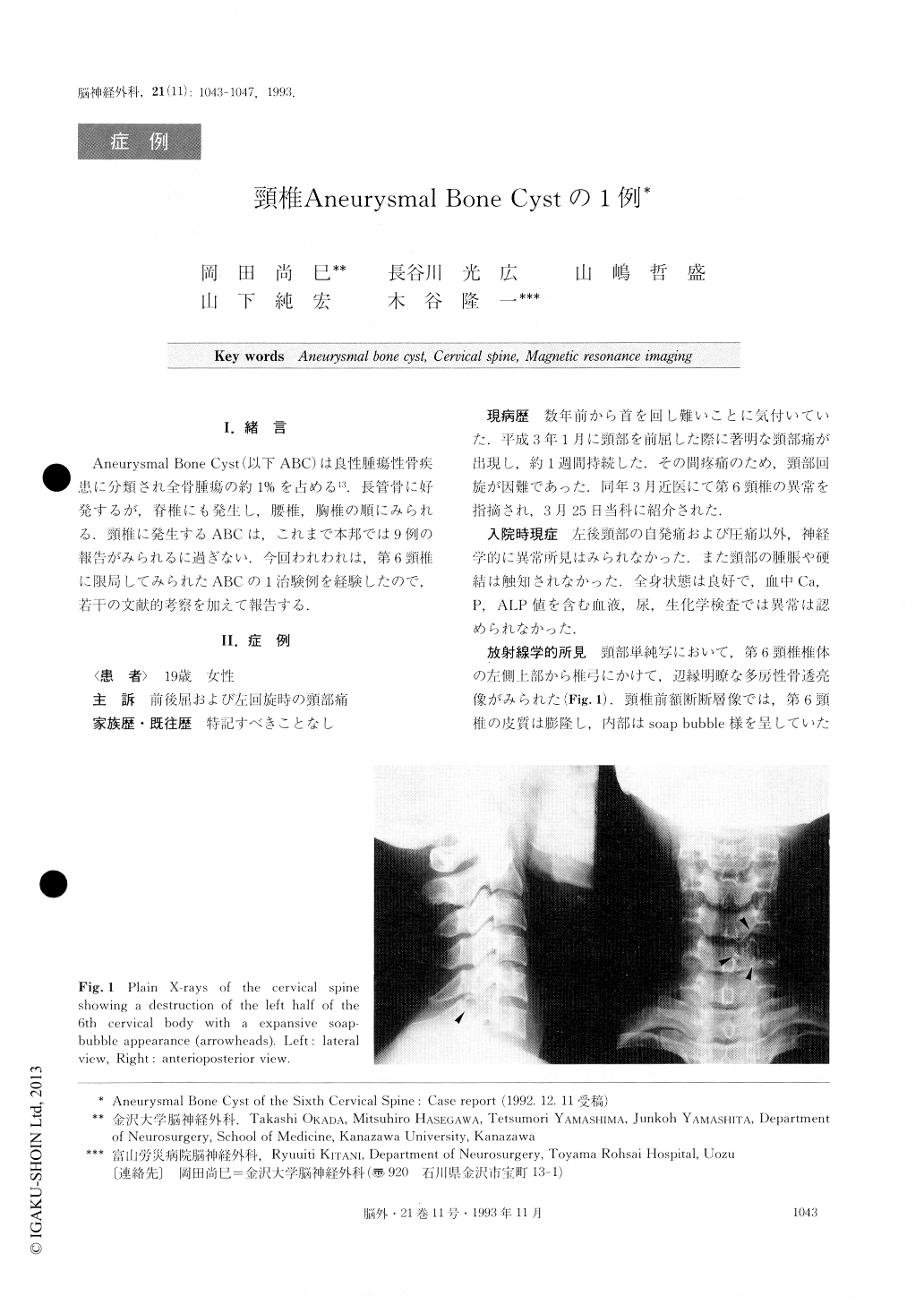
A 19-year-old girl was admitted with a history of diffi-culty in moving her neck for several years and a sudden onset of neck pain three months before. Plain radio-graphs of the cervical spine revealed destruction of the left half of the 6th cervical body with an expansive soap-bubble appearance.
Neurological examination on admission was within normal limits. The angiography and bone scintigraphy revealed no abnormality. MRI of T1-weighted image showed a cystic lesion with various signal intensities.

Copyright © 1993, Igaku-Shoin Ltd. All rights reserved.


