Japanese
English
- 有料閲覧
- Abstract 文献概要
- 1ページ目 Look Inside
I.はじめに
脊髄硬膜外嚢胞は1934年Elsbergら1)により初めて臨床像が明らかにされた稀な疾患である.青少年期の胸椎に好発し緩徐進行性の神経症状を示すが,ほぼ30%の例で症状の増悪緩解がみられる4,9).われわれは少年期の両下肢麻痺発症から長い緩解期を経て42歳の時にようやく診断が確定し外科的治療を行った1例を経験したが貴重な症例と考えられるので報告する.
We reported a peculiar case of a patient with a thoracic extradural cyst, who had a 30-year history of thoracic myelopathy with a very long-term remission.
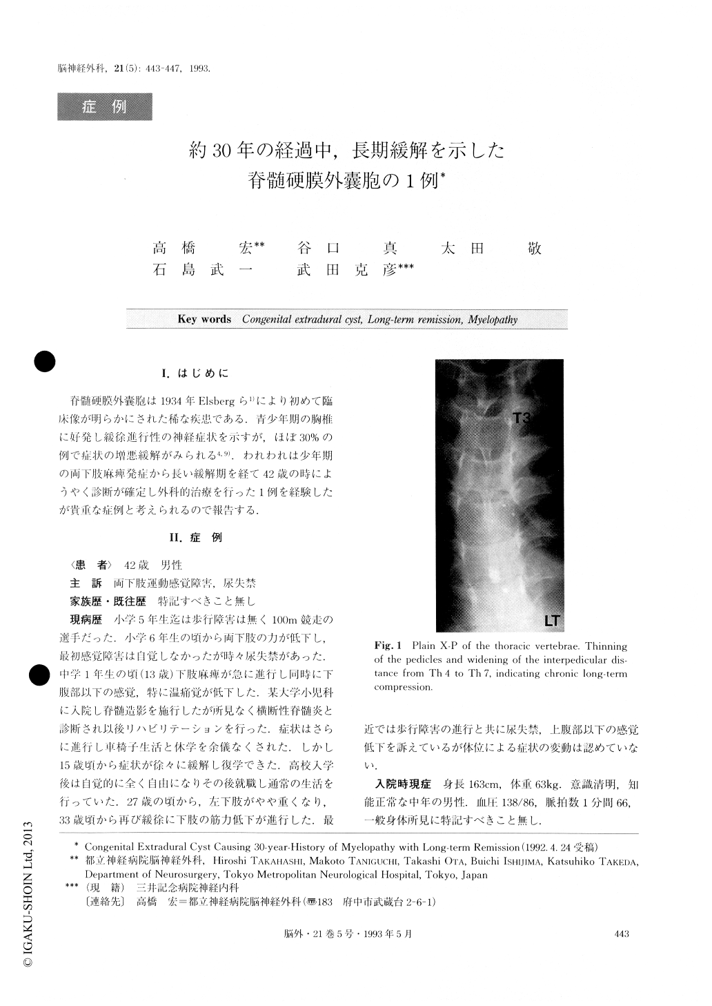
A 42-year-old male was admitted to our hospital complaining of gait disturbance and hypesthesia of his bilateral lower limbs. Neurological examination revealed that he had spastic paraparesis and hypesthesia of all sensory modalities below Thll/12 dermatomes. Investigation of his past history disclosed that he had experienced gait disturbance due to paraplegia 29 years previously and had been able to move only with the aid of wheel-chairs. However these signs had resolved themselves gradually in a period of 3 years, and after that he had been able to live almost without any neurological handicaps until the recent appearance of weakness in both of his legs. Neuroradiological study revealed that plain X-Ps of the thoracic spine showed thinning of the pedicles and widening of the interpedicular distances between Th4-Th7 vertebrae. The old iodine contrast media injected 29 years previously for myelography in an other hospital was also observed. MRI showed a dorsal cystic mass compressing the thoracic spinal cord and subarachnoid space. Myelography showed also that the spinal cord and subarachnoid space were compressed anteriorly between the level of Th3 to Th7 by the dorsal mass.
CT myelography disclosed that soon after the injection of contrast media a small part of the cyst was visualized as having direct communication with the subarachnoid space. The contrast media penetrated the major part of the cyst only 19 hours after its injection. With these observations a thoracic extradural cyst causing exacerbation after long-term remission was diagnosed.
Operation disclosed that the extradural cyst was composed of tough dura mater-like wall externally and arachnoid-like thin membranes internally. The cyst was removed totally with the old contrast media still trapped inside it. The post-operative course was uneventful. Although the patient still had slight weakness of the left lower limb and hypesthesia of the right lower limb, he was able to return to his previous occupation.
Copyright © 1993, Igaku-Shoin Ltd. All rights reserved.