Japanese
English
- 有料閲覧
- Abstract 文献概要
- 1ページ目 Look Inside
I.はじめに
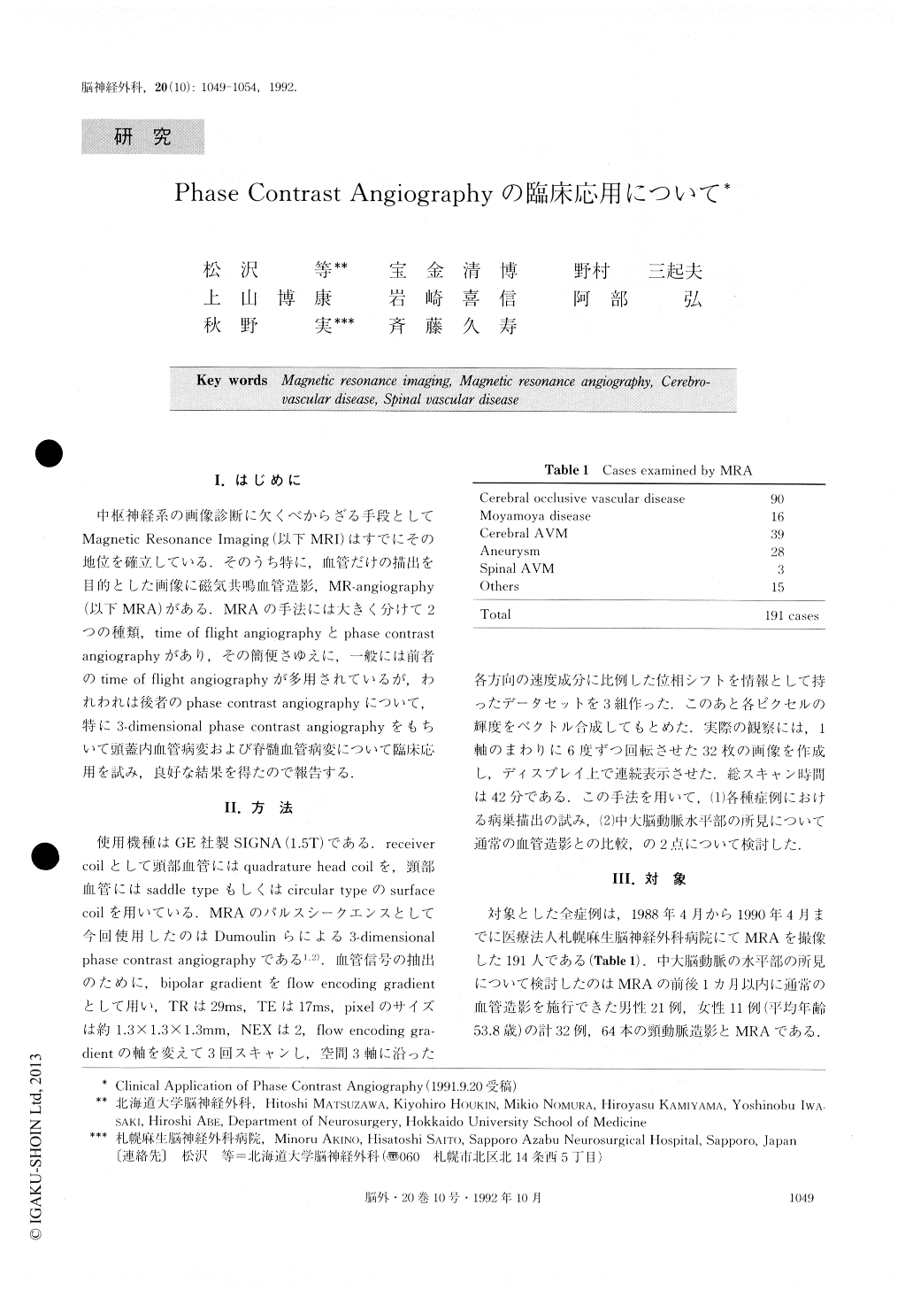
中枢神経系の画像診断に欠くべからざる手段としてMagnetic Resonance Imaging(以下MRI)はすでにその地位を確立している.そのうち特に,血管だけの描出を目的とした画像に磁気共鳴血管造影,MR-angiography(以下MRA)がある.MRAの手法には大きく分けて2つの種類,time of flight angiographyとphase contrastangiographyがあり,その簡便さゆえに,一般には前者のtime of flight angiographyが多用されているが,われわれは後者のphase contrast angiographyについて,特に3—dimensional phase contrast angiographyをもちいて頭蓋内血管病変および脊髄血管病変について臨床応用を試み,良好な結果を得たので報告する.
“Phase Contrast Angiography” is a new technique of Magnetic Resonance Angiography as reported by Dumoulin CL et al. Using this technique, we can obtain images of vessels (angiograms) without injection of con-trast medium.
We present the results of phase contrast angiography on cerebral and spinal vascular disease.
We utilized the General Electric SIGNA, 1.5 tesla NMR. One hundred and ninety-one cases of cerebral and spinal vascular diseases were scanned using phase con-trast angiography. Included were 90 cases of occlusive vascular disease, 16 cases of moyamoya disease, 39 cases of arteriovenous malformation, and 28 aneurysms. The phase contrast angiography uses flow encoding gradient pulses, which impart a velocity-dependent phase shift to the transverse magnetization of moving spins. The re-sulting image contains only information from the moving spins ; while information from stationary tissue remains suppressed. In cases using 3-D angiogram, we made 32images 6 degrees apart in their projection direction and displayed them on a video terminal.
We were able to visualize occlusions of vessels, aneurysms, bypassed vessels, and abnormal vessels of arteriovenous malformations. Retrospective evaluation comparing phase contrast angiography with convention-al angiography of the stenotic findings on the horizontal portion of middle cerebral arteries (64 vessels of 32 pa-tients), resulted in a false positive ratio of 39.1%. We obtained clinically valuable results regarding the hydrodynamics of patients using “phase contrast angiography” non-invasively. These results reveal not only “anatomical” images of vessels, but also “function-al” images, which are sensitive to the pattern of the blood flow. This study would strongly suggest that phase contrast angiography presents a valuable tool for the clinical diagnosis of cerebral and spinal vascular dis-eases.

Copyright © 1992, Igaku-Shoin Ltd. All rights reserved.


