Japanese
English
- 有料閲覧
- Abstract 文献概要
- 1ページ目 Look Inside
I.はじめに
小脳橋角槽の第7・8脳神経と周囲動脈は複雑な走行を示す.近年,顔面けいれんやめまい・耳鳴り患者への神経減圧術の普及や早期に発見されるintracanalicularacoustic neurinoma手術例の増加とともに,内耳孔近傍の微小外科解剖特に顔面・聴神経束と周囲動脈の外科解剖の重要性が更に増加してきている.この部の顔面・聴神経を栄養する動脈を損傷すると,術後難聴や顔面神経麻痺を生じることがあり,術中正確なオリエンテーションをつけることが大切である.
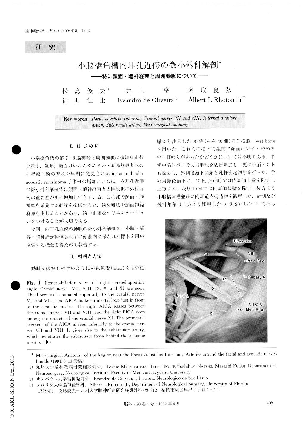
今回,内耳孔近傍の動脈の微小外科解剖を,小脳・脳幹・脳神経が損傷されずに頭蓋内に保たれた標本を用い検索する機会を得たので報告する.
The microsurgical anatomy of the cerebellopontine cistern around the porus acusticus was studied under the surgical microscope, using the heads of 20 cadavers. The relationships among the porus acusticus, facial and acoustic nerves, and neighboring arteries were noted carefully. The arteries were the meatal loop of the cerebellar artery, the internal auditory artery (I. A. A.), the subarcuate artery (S. A.), and the perforating artery (P. A.). The cerebellar arteries made the meatal loop and the arterialnerve complex while they passed in front of the porus acusticus. Most of the cerebellar arteries were the main or rostral trunk of the AICA. The I. A. A. supplying the facial and acoustic nerves usually originated from the meatal segment of the cerebellar artery and ran into the anterior part of the porus acusticus to enter the internal auditory canal. The S. A. penetrating the subarcuate fossa made a common trunk with I. A. A. or branched from the cerebellosubarcuate artery. One recurrent P. A., a special type of the P. A., was usually present on each side. Since the artery ran between the nerve bundles of the facial and acoustic nerves, it could not be seen through the lateral suboccipital approach.

Copyright © 1992, Igaku-Shoin Ltd. All rights reserved.


