Japanese
English
- 有料閲覧
- Abstract 文献概要
- 1ページ目 Look Inside
I.はじめに
Meningioangiomatosisは,Worster-Droughtら15)に提唱されたhamartomaの性格の強い病態であり,その大部分がvon Reckhnghausen病に合併する疾患である.von Recklinghausen病に合併しないmeningioangioma—tosisの症f列はKasantikulら21)の報告にはじまるが,現在まで11例が報告されている.われわれは,von Reck—linghausen病に合併していないと考えられるMeningio—angiomatosisの乳児例を経験したので神経放射線学的特徴及び病態に関して,若干の文献的考察を加えて報告する.
Abstract
We report here a rare case of meningioangiomatosis in an infant, not associated with von Recklinghausen's disease.
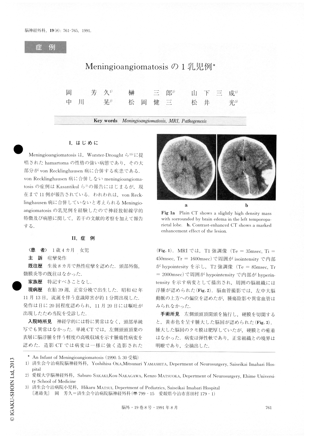
A 14-month-old female was admitted because of sei-zures. Neurological findings on admission were normal. Computed tomography showed a slightly high densitymass with marked contrast enhancement in the left temporoparietal lobe. Magnetic resonance image (MRI) revealed a sightly hypointensive lesion surrounded by an isointensive band on T1-weighted image, and a hy-perintensive lesion surrounded by a slightly hypointen-sive band on T2-weighted image. Brain edema was shown to a certain extent around the lesion on MRI. Left carotid angiography demonstrated a slightly up-ward shift of the left middle cerebral artery, but no abnormal vascularity was shown.
A temporoparietal craniotomy was performed. A yel-lowish red, elastic soft tumor was observed in the left temporal lobe. The tumor resembled hyperemic hyper-trophic gyri and was well demarcated. Total removal of the tumor was performed.
Pathological diagnosis was meningioangiomatosis. The patient is still doing well 3 years and 5 months after the operation. There was no evidence of recur-rence on computed tomography at the 3-year follow up. She didn't have any stigmata, such as cafe au lait spots or neurofibromas suggesting von Recklinghausen's dis-ease.

Copyright © 1991, Igaku-Shoin Ltd. All rights reserved.


