Japanese
English
- 有料閲覧
- Abstract 文献概要
- 1ページ目 Look Inside
I.はじめに
Hypertrophic Pachymeningitis(以下HP)は1869年Charcotら4)により初めて記載された硬膜の慢性炎症性肥厚を特徴とする比較的稀な疾患であり,頸部,胸部の脊髄神経根及び脊髄の圧迫症状を早することが多いとされている10,24).今回われわれは水頭症,脳神経症状,小脳症状などの頭蓋内症状を主体としたHypertrophicCranial Pachymeningitis(以下HCP)の1例を経験したので若干の文献的考察を加え報告する.
Abstract
A case of hypertrophic cranial pachymeningitis was reported.
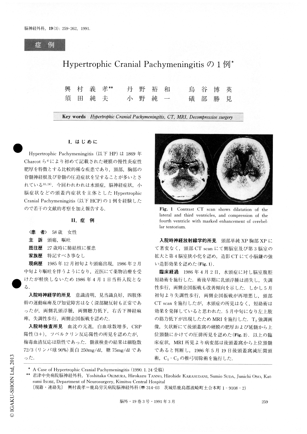
A 58-year-old female presented the symptoms of headache and vomiting. At the age of 27, she had suf-fered from tuberculosis. Neurological examination on admission revealed bilateral papilledema, bilateral hear-ing disturbance, right hypoglossal nerve palsy, ataxic gait, and bilateral intentional tremor. CT scan showed dilatation of the lateral and third ventricles, and com-pression of the fourth ventricle with marked enhance-ment of cerebellar tentorium. A ventriculoperitoneal shunt was installed bringing about improvement in bi-lateral papilledema, ataxic gait, and bilateral intentional tremor. One month later, ataxic gait and bilateral inten-tional tremor recurred, and monoparesis of the left up-per extremity developed. MRI demonstrated hyper-trophic dura mater in the posterior fossa and compress-ed cervical spinal cord. Decompressive surgery was per-formed bringing about remarkable clinical improve-ment. The pathological specimen showed thickening of the dura mater with concentric layers of dense fibrous tissue infiltrated with plasma cells. A diagnosis of hypertrophic cranial pachymeningitis was established. Three years later, the clinical features were found un-changed, but contrast enhancement of cerebellar tentor-ium had progressed markedly.
Hypertrophic pachymeningitis is a uncommon dis-ease. But it should be noted that intracranial involve-ment is very rare. The etiology, symptomatology, neuroradiology, and treatment are discussed and the literature is reviewed.

Copyright © 1991, Igaku-Shoin Ltd. All rights reserved.


