Japanese
English
- 有料閲覧
- Abstract 文献概要
- 1ページ目 Look Inside
- 参考文献 Reference
Point
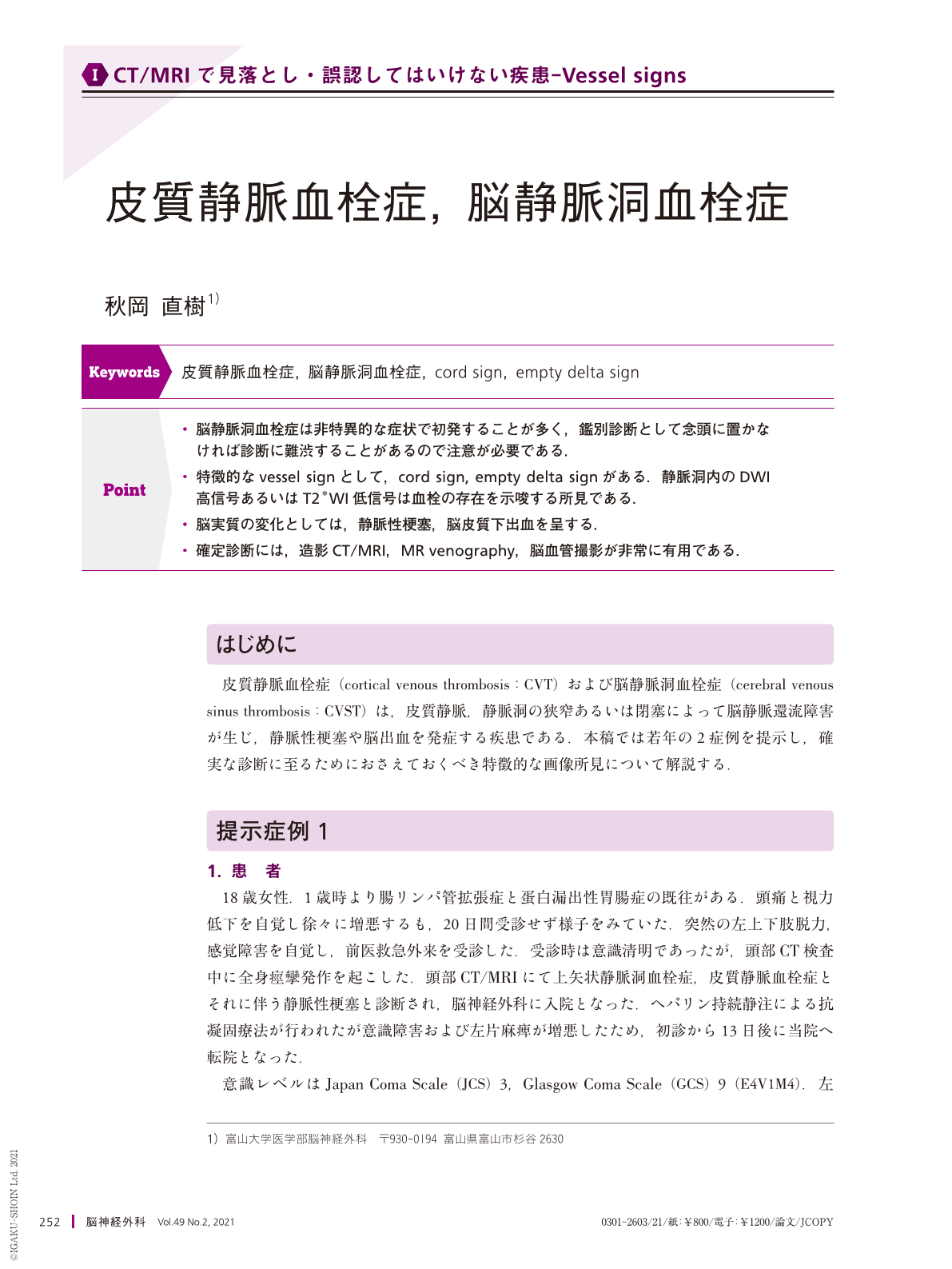
・脳静脈洞血栓症は非特異的な症状で初発することが多く,鑑別診断として念頭に置かなければ診断に難渋することがあるので注意が必要である.
・特徴的なvessel signとして,cord sign, empty delta signがある.静脈洞内のDWI高信号あるいはT2*WI低信号は血栓の存在を示唆する所見である.
・脳実質の変化としては,静脈性梗塞,脳皮質下出血を呈する.
・確定診断には,造影CT/MRI,MR venography,脳血管撮影が非常に有用である.
The author reports the cases of two young patients with cortical venous thrombosis(CVT)and cerebral venous sinus thrombosis(CVST)and demonstrates that CT and MRI investigations are critical for the diagnosis. The first case was an 18-year-old woman who developed symptoms of intracranial hypertension and, 20 days later, suffered from left hemiparesis and generalized seizures. A plain CT scan revealed an increased density of cortical veins(“cord sign”). MR venography revealed superior sagittal sinus thrombosis and right frontal lobe cortical vein thrombosis. The second case was a 15-year-old boy who developed intracranial hypertension symptoms and went to various hospitals but the symptoms progressed. Six weeks after the onset, contrast-enhanced MRI showed an “empty delta sign” and superior sagittal sinus thrombosis was diagnosed. The patients had a history of protein-losing enteropathy and Behçet's disease. Both patients were treated with endovascular and anticoagulation therapy while also treating the primary disease, resulting in thrombus reduction in the sinus and good outcomes. CVT and CVST can lead to severe conditions, so a full understanding of the imaging findings is essential for an accurate diagnosis.

Copyright © 2021, Igaku-Shoin Ltd. All rights reserved.


