Japanese
English
- 有料閲覧
- Abstract 文献概要
- 1ページ目 Look Inside
- 参考文献 Reference
Ⅰ.緒 言
低髄液圧症・脳脊髄液漏出症は,そのほとんどが脊髄レベルからの脳脊髄液の漏出によって髄液圧低下や液量減少を来す病態と考えられている10,14).これらは,国際頭痛分類第3版beta版12)(以下,ICHD-3β)の「7.2低髄液圧による頭痛」に含まれる疾患と考えられ,「厚生労働省 脳脊髄液減少症の診断・治療法の確立に関する研究班」(以下,厚労省研究班)により画像診断基準が出された13).特に低髄液圧症では,病初期に起立に伴い増悪傾向のある頭痛(起立性頭痛)が出現し,経過中の一定の時期に頭部造影MRIにおいてびまん性硬膜肥厚をはじめとする特徴的な所見を認める場合が多く,腰椎部髄液圧測定では60mmH2O以下の低髄液圧を確認できることが多い13,14).しかし最近の症例の集積により,特徴的な画像所見を認めない症例や正常髄液圧を示す症例もあることが判明してきており,“髄液漏れ”が存在しても,必ずしも異常検査所見を認めるとは限らない6,12,14).
ICHD-3βでは,画像検査によって髄液漏出の証拠を示すべきとされ12),現時点では,脊髄MRI/MR myelography(以下,脊髄MRI)・CT myelography(以下,CTM)・radioisotope cisternography(以下,RIC)などの検査が行われる13).後二者は腰椎穿刺を必要とすることから,脊髄MRIを優先して施行すべきである13).脊髄MRI横断像にて,硬膜外腔に水貯留(以下,硬膜外水腫)所見を認める場合があり,髄液漏出の存在を示唆すると考えられる4,5).本所見に対してはfloating dural sac sign(FDSS)という名称が提唱されている4).今回,低髄液圧症と診断した症例群の診断治療経過と脊髄硬膜外水腫所見について考察する.
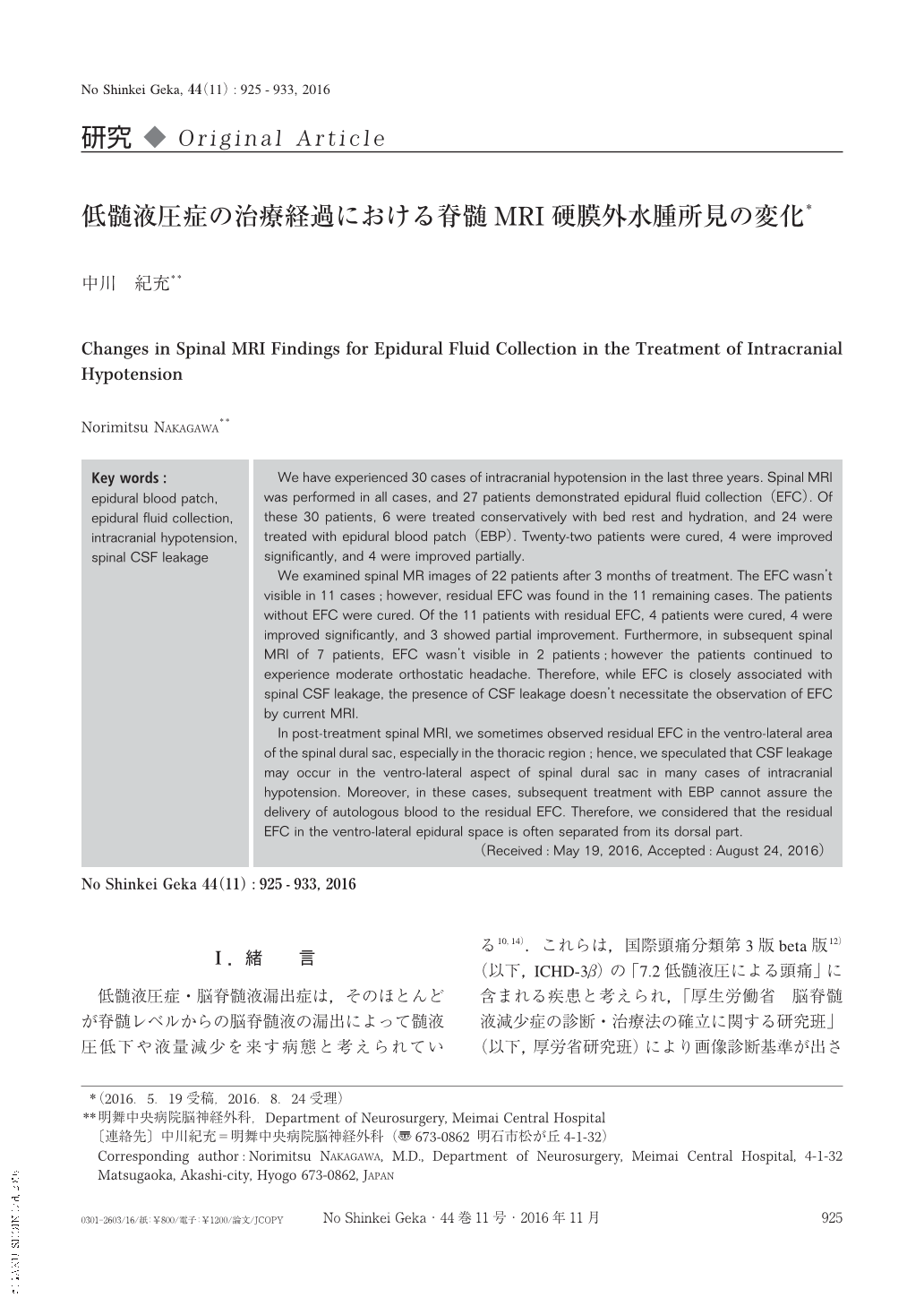
We have experienced 30 cases of intracranial hypotension in the last three years. Spinal MRI was performed in all cases, and 27 patients demonstrated epidural fluid collection(EFC). Of these 30 patients, 6 were treated conservatively with bed rest and hydration, and 24 were treated with epidural blood patch(EBP). Twenty-two patients were cured, 4 were improved significantly, and 4 were improved partially.
We examined spinal MR images of 22 patients after 3 months of treatment. The EFC wasn't visible in 11 cases;however, residual EFC was found in the 11 remaining cases. The patients without EFC were cured. Of the 11 patients with residual EFC, 4 patients were cured, 4 were improved significantly, and 3 showed partial improvement. Furthermore, in subsequent spinal MRI of 7 patients, EFC wasn't visible in 2 patients;however the patients continued to experience moderate orthostatic headache. Therefore, while EFC is closely associated with spinal CSF leakage, the presence of CSF leakage doesn't necessitate the observation of EFC by current MRI.
In post-treatment spinal MRI, we sometimes observed residual EFC in the ventro-lateral area of the spinal dural sac, especially in the thoracic region;hence, we speculated that CSF leakage may occur in the ventro-lateral aspect of spinal dural sac in many cases of intracranial hypotension. Moreover, in these cases, subsequent treatment with EBP cannot assure the delivery of autologous blood to the residual EFC. Therefore, we considered that the residual EFC in the ventro-lateral epidural space is often separated from its dorsal part.

Copyright © 2016, Igaku-Shoin Ltd. All rights reserved.


