Japanese
English
- 有料閲覧
- Abstract 文献概要
- 1ページ目 Look Inside
I.はじめに
遅発性放射線脳壊死(以下radiation necrosis)は,まず血管壁が直接障害を受けてフィブリノイド変性を生じ,神経組織に対する障害はその結果からくる二次的なものであるという考え方が大方の見解である.しかし,新生物でないにもかかわらず,高度の脳浮腫を伴ったmass effectがなぜ持続するのかなど,その発生機序については不明な点が多い.
われわれはすでに前報でgliomaにおける脳浮腫の発生機序を血管透過性の面から検討を加え報告した8,9).微小血管の面から見ると,前者は正常脳血管の放射線による変性であり,後者は腫瘍の新生血管である.そこでradiation necrosisの微小血管の形態学的変化を電顕的に検索し,脳浮腫発生の一因としての血管透過性について,glioma(glioblastoma, astrocytoma)と対比して検討を加え,興味ある所見が得られたので報告する.
In a patient with a delayed cerebral radionecrosis, we examined the ultrastructural changes of capillaries and discussed the cause of the development of severe brainedema which is one of specific features of the radionec-rosis.
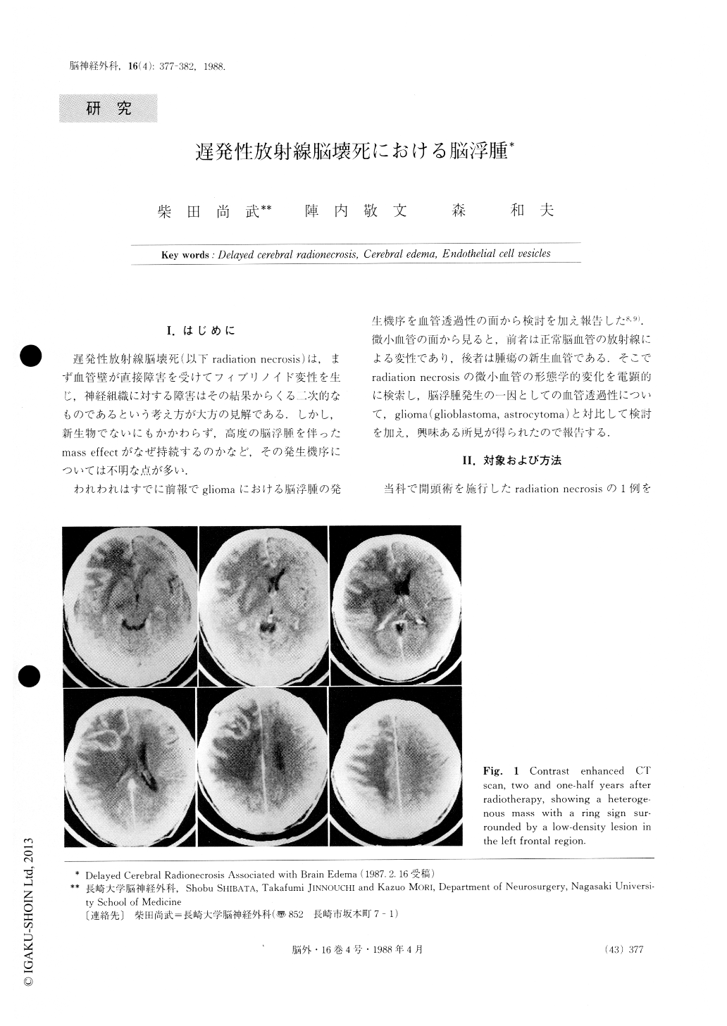
A 42-year-old male was found to have cerebral radionecrosis two and one-half years following split-course radiotherapy which was done after subtotal ex-cision of a left parasagittal meningioma. The surgical specimens were studied with conventional ultrathin sec-tion and freeze-fracture replica techniques.

Copyright © 1988, Igaku-Shoin Ltd. All rights reserved.


