Japanese
English
- 有料閲覧
- Abstract 文献概要
- 1ページ目 Look Inside
I.はじめに
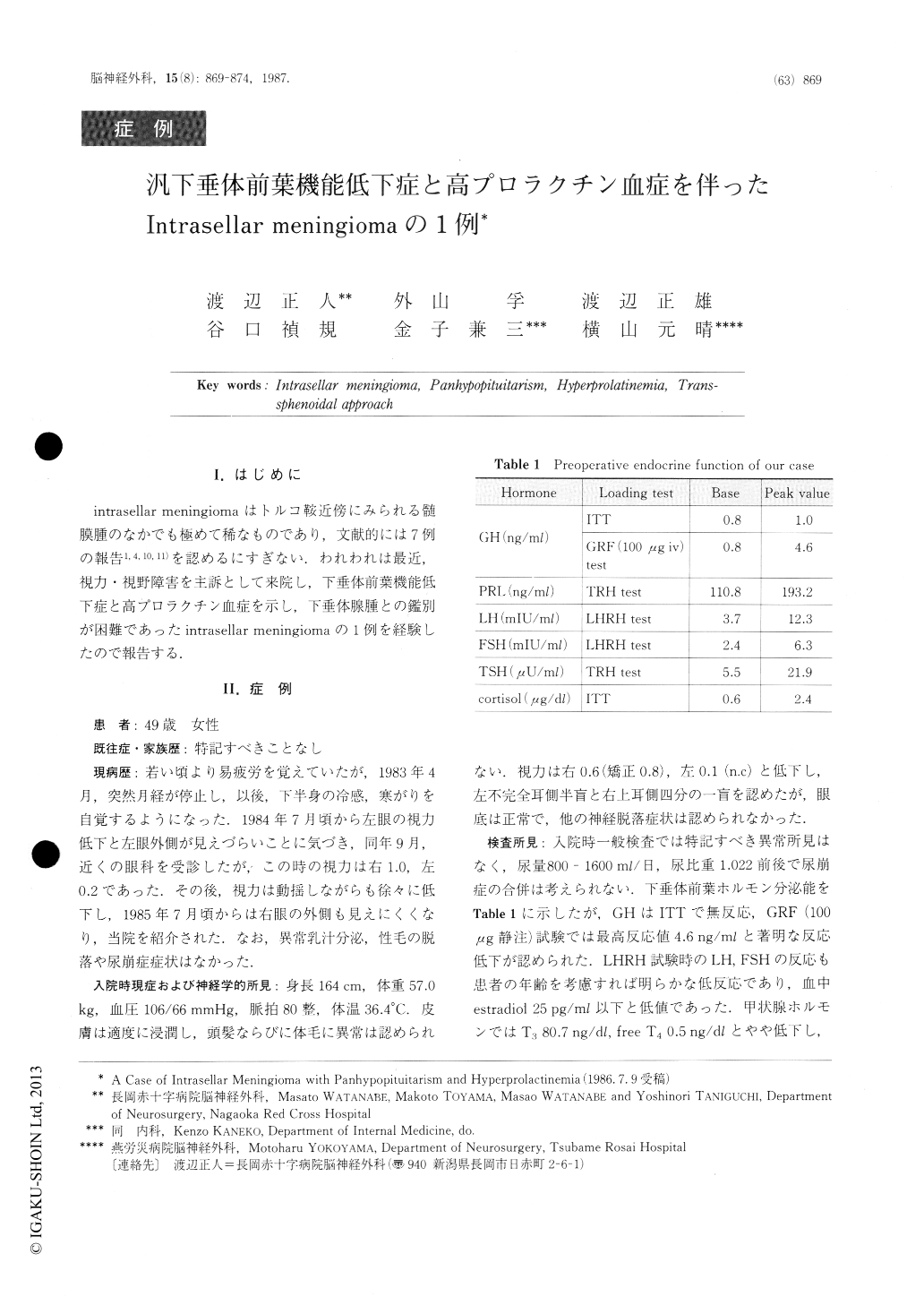
intrasellar meningiomaはトルコ鞍近傍にみられる髄膜腫のなかでも極めて稀なものであり,文献的には7例の報告1,4,10,11)を認めるにすぎない.われわれは最近,視力・視野障害を主訴として来院し,下垂体前葉機能低下症と高プロラクチン血症を示し,下垂体腺腫との鑑別が困難であったintrasellar meningiomaの1例を経験したので報告する.
A case of intrasellar meningioma is reported. A 49-year-old woman was admitted to our hospital on July 22, 1985, complaining of reduced visual acuity and visual field defect. Visual acuity was 0.6 in the right eye and 0.1 in the left eye. Visual field examination re-vealed upper temporal quadrantanopsia on the right side and incomplete temporal hemianopsia on the left side. Ocular fundi were normal. X-ray films of the skull showed a balloon-shaped sella turcica with "double floor". CT scan showed a isodense mass with central low density occupying the intrasellar and suprasellar re-gion.

Copyright © 1987, Igaku-Shoin Ltd. All rights reserved.


