Japanese
English
- 有料閲覧
- Abstract 文献概要
- 1ページ目 Look Inside
I.はじめに
慢性硬膜下血腫(特にisodensityなもの)の診断における脳血管CT(cerebral computed angiotomogra—phy)の有用性については既に報告してきた1,15,16).脳血管CTはこのほか,脳血管病変自体のスクリーニング法としても有用である2,7,8,15,16).最近われわれは,手技も簡便で,技術的にも進歩した「脳血管CT超高速度撮影法(ultrafast cerebral computed angiotomogra—phy)」を開発し8,16),外来診療に応用している.今回は本法を用いて,慢性硬膜下血腫の部位別,確定診断を行い,同時に潜在性未破裂脳動脈瘤の合併を術前に診断し得た1症例を経験したので報告する.
One case of chronic subdural hematoma associated with an unruptured cerebral aneurysm detected by cerebral computed angiotomography is reported.
A 14-year-old female slipped and hit her head without loss of consciousness, one month ago. Re-cently she complained of headaches and visited the department of Neurosurgery, Washokai Sadamoto Hospital on May 21, 1985. There were no physical and neurological signs on examination.
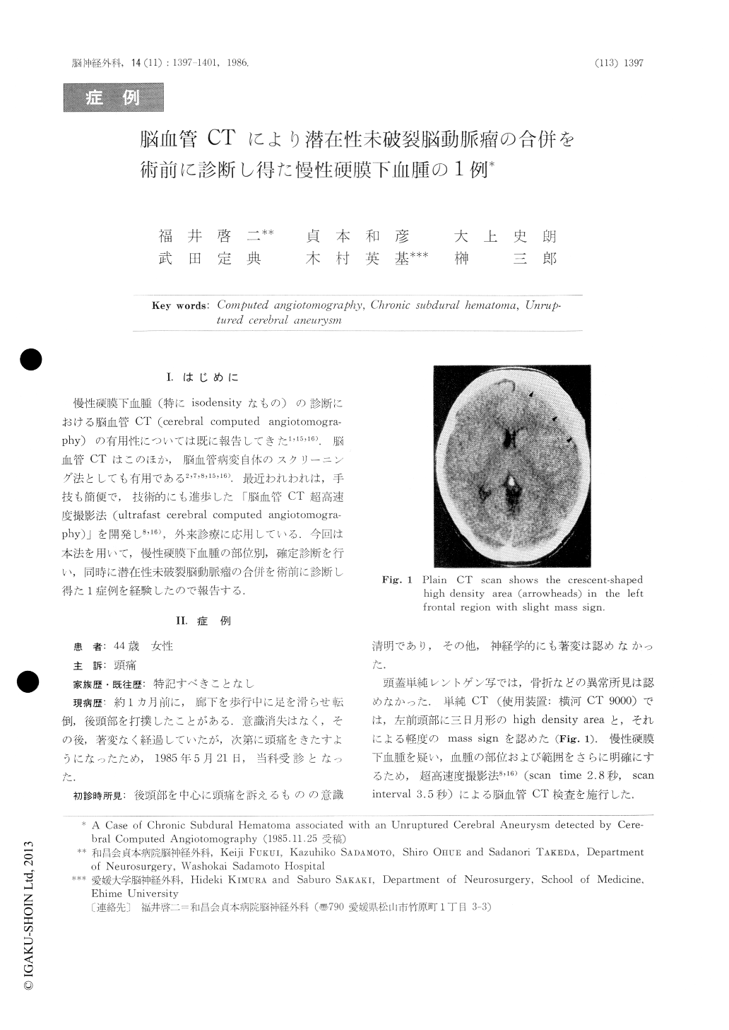
Plain CT scans showed a crescent-shaped high density area in the left frontal region with a slight mass sign.

Copyright © 1986, Igaku-Shoin Ltd. All rights reserved.


