Japanese
English
- 有料閲覧
- Abstract 文献概要
- 1ページ目 Look Inside
I.はじめに
chondromyxoid fibromaは,1948年,JaffeおよびLichtenstein15)により命名された軟骨組織に山来する良性腫瘍であり,その頻度は骨腫瘍全体の1%以下と稀な疾患である23).発生部位は脛骨,腓骨,大腿骨など長管骨が大部分を占め,頭蓋骨発生例は,われわれの渉猟しえた範囲ではこれまで9例の報告をみるにすぎない,最近われわれは,前頭骨に発生したchodromyxid fibro-ma の1症例を経験したので,若干の文献的考察を加え報告する.
A case of chondromyxoid fibroma of the skull isreported.
A 20-year-old boy visited our clinic on December,1982 because of a recurrent forehead tumor. He hada 4×4×1.5cm tumor on the right side of foreheadand a 3×3×0.5cm tumor on the left.
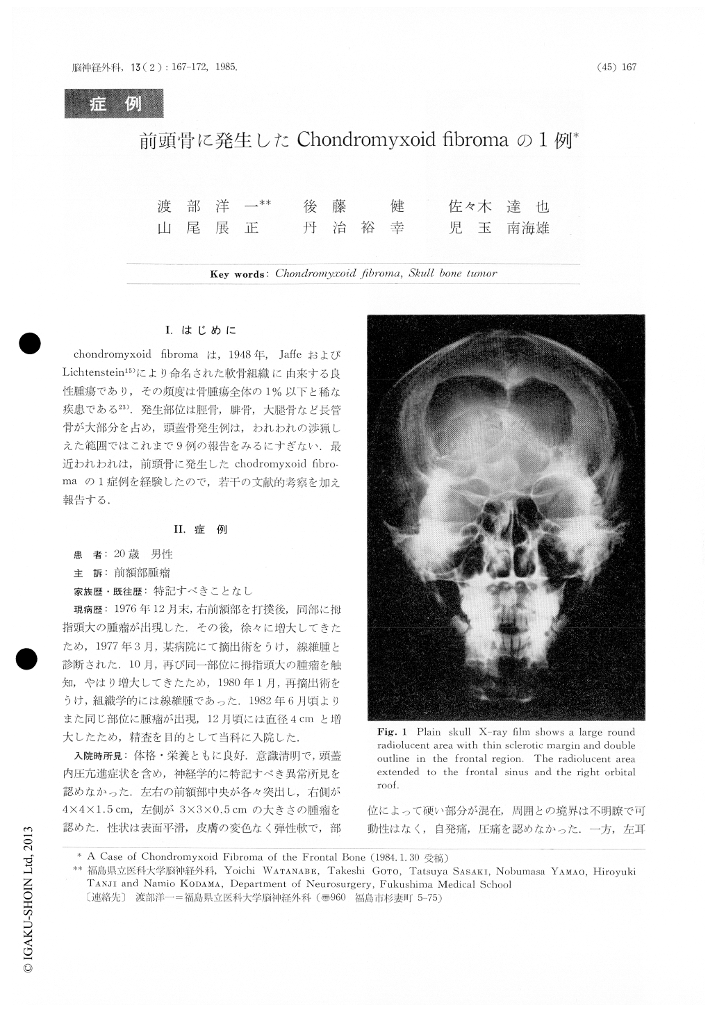
Neurological examination showed no abnormalities.Skull X-ray film showed a large round radiolucentarea with clear sclerotic margin in the frontal boneand right orbit. Right carotid angiogram showedmarked posterior displacement of the anterior cerebralartery, but no tumor stain. Plain CT scan showed amass with iso to low density area in the frontal region.

Copyright © 1985, Igaku-Shoin Ltd. All rights reserved.


