Japanese
English
- 有料閲覧
- Abstract 文献概要
- 1ページ目 Look Inside
Ⅰ.はじめに
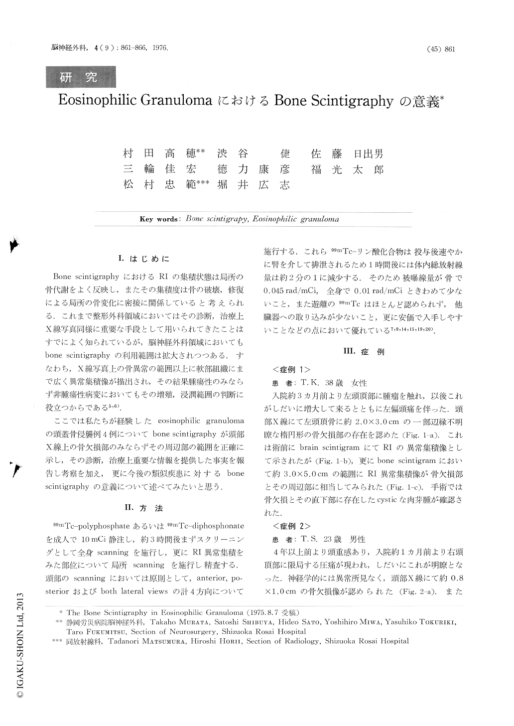
Bone scintigraphyにおけるRIの集積状態は局所の骨代謝をよく反映し,またその集積度は骨の破壊,修復による局所の骨変化に密接に関係していると考えられる.これまで整形外科領域においてはその診断,治療上X線写真同様に重要な手段として用いられてきたことはすでによく知られているが,脳神経外科領域においてもbone scintigraphyの利用範囲は拡大されつつある.すなわち,X線写真上の骨異常の範囲以上に軟部組織にまで広く異常集積像が描出され,その結果腫瘍性のみならず非腫瘍性病変においてもその増殖,浸潤範囲の判断に役立つからである5,6).
ここでは私たちが経験したeosinophilic granulomaの頭蓋骨侵襲例4例についてbone scintigraphyが頭部X線上の骨欠損部のみならずその周辺部の範囲を正確に示し,その診断,治療上重要な情報を提供した事実を報告し考察を加え,更に今後の類似疾患に対するbone scintigraphyの意義について述べてみたいと思う.
The significance of bone scintigraphy using 99mTc-polyphosphate or 99mTc-diphosphate in the diagnosis of eosinophilic granuloma was discussed on the basis of our experience with four cases.
The summary is following;
1. On diagnostic procedure of eosinophilic granuloma, it was found that abnormal RI accumulation in the bone scintigram was more marked than that in the ordinary brain scintigram.
2. The bone scintigram of eosinophilic granuloma showed more accurately the extent of skull involvement of cellular infiltration than the plain skull X-rays or brain scintigram.

Copyright © 1976, Igaku-Shoin Ltd. All rights reserved.


