Japanese
English
- 有料閲覧
- Abstract 文献概要
- 1ページ目 Look Inside
Ⅰ.序文
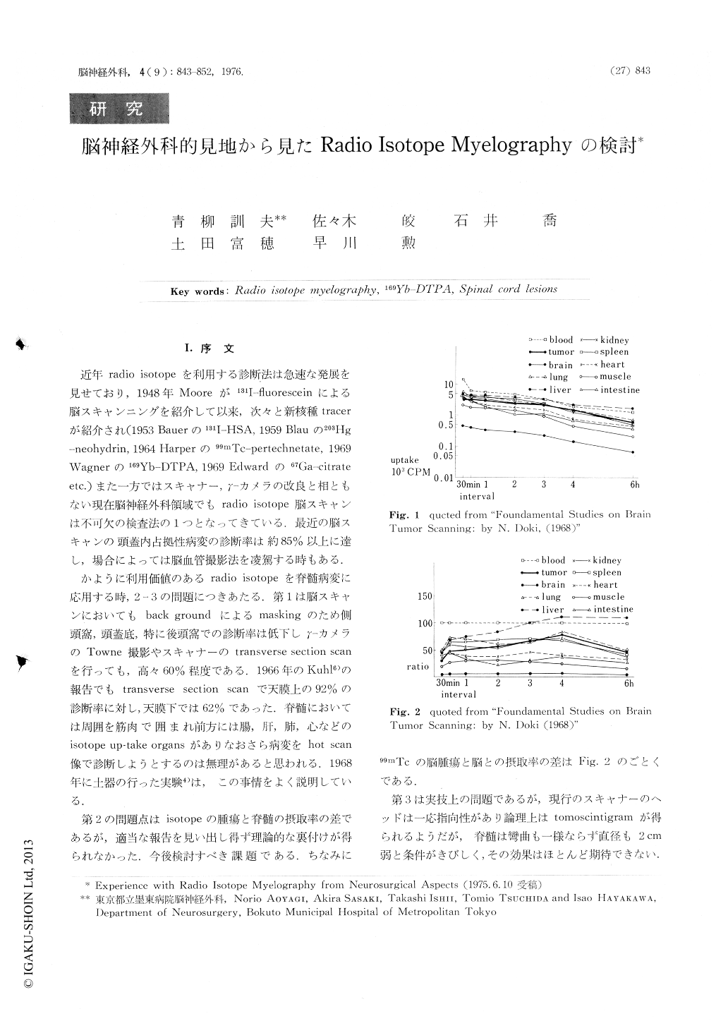
近年radio isotopeを利用する診断法は急速な発展を見せており,1948年Mooreが131I-fluoresceinによる脳スキャンニングを紹介して以来,次々と新核種tracerが紹介され(1953 Bauerの131I-HSA,1959 Blauの203Hg-neohydrin, 1964 Harperの99mTc-pertechnetate,1969 Wagnerの169Yb-DTPA,1969 Edwardの67Ga-citrate etc.)また一方ではスキャナー,γ-カメラの改良と相ともない現在脳神経外科領域でもradio isotope脳スキャンは不可欠の検査法の1つとなってきている,最近の脳スキャンの頭蓋内占拠性病変の診断率は約85%以上に達し,場合によっては脳血管撮影法を凌駕する時もある.
かように利用価値のあるradio isotopeを脊髄病変に応用する時,2-3の問題につきあたる.第1は脳スキャンにおいてもback groundによるmaskingのため側頭窩,頭蓋底,特に後頭窩での診断率は低下しγ-カメラのTowne撮影やスキャナーのtransverse section scanを行っても,高々60%程度である.1966年のKuhl6)の報告でもtransverse section scanで天膜上の92%の診断率に対し,天膜下では62%であった.
Experience with Radio Isotope Myelography is reported here. 169Yb-DTPA as a tracer was intrathecally injected at lumbar region in twenty three patients with various spinal cord lesions. The first scanning is perfomed after comfirming by γ-camera that the tracer reaches to the lesion, the second and the third scannings are clone according to the ascending rate of the tracer.
(I) Normal scintimyelogram
(A) In normal case, the shape of the Radio Isotope Myelogram well corresponds the shape of anatomical suharachnoid space.
(B) In normal adult cases, the tracer comes up to the cisterna magna in 20-25 minutes after the lumbar injection.

Copyright © 1976, Igaku-Shoin Ltd. All rights reserved.


