Japanese
English
- 有料閲覧
- Abstract 文献概要
- 1ページ目 Look Inside
- 参考文献 Reference
Ⅰ.はじめに
脳神経外科領域におけるインドシアニングリーン(indocyanine green;ICG)の螢光特性を利用した術中螢光血管造影の有用性がRaabeら4)により報告されて以降,浅側頭動脈─中大脳動脈吻合術6),脳動脈瘤クリッピング術3)などに有用性が報告されている.ICG videoangiography(ICG-VA)は,静脈注射のみで施行可能であり,短時間で評価できるため今後もますます発展していくと思われるが,頚動脈内膜剝離術における有用性を報告した文献は未だ少数である2,6).
頚動脈内膜剝離術(carotid endarterectomy:CEA)においては,内頚動脈遠位部をプラークの存在しない部位まで十分剝離することが重要であるが,外表からの観察でプラークの遠位端を正確に同定することは困難である.われわれは動脈切開前にICG-VAを行い,プラーク遠位端を確認し得たので,その有用性およびpitfallを報告する.
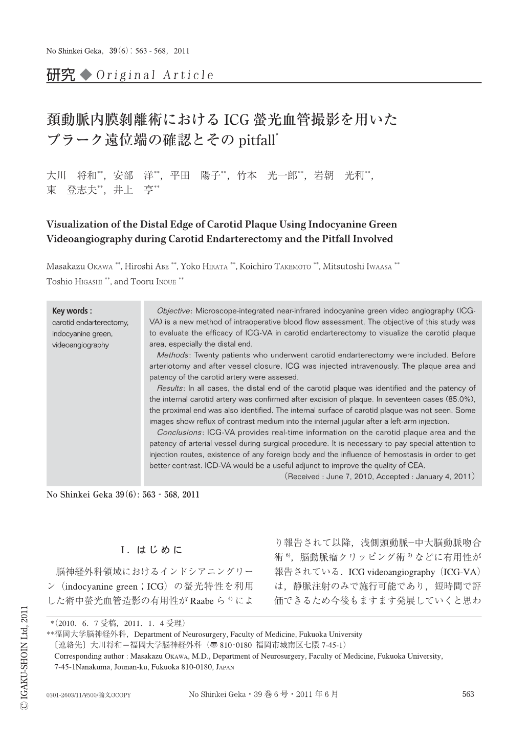
Objective: Microscope-integrated near-infrared indocyanine green video angiography (ICG-VA) is a new method of intraoperative blood flow assessment. The objective of this study was to evaluate the efficacy of ICG-VA in carotid endarterectomy to visualize the carotid plaque area,especially the distal end.
Methods: Twenty patients who underwent carotid endarterectomy were included. Before arteriotomy and after vessel closure, ICG was injected intravenously. The plaque area and patency of the carotid artery were assesed.
Results: In all cases, the distal end of the carotid plaque was identified and the patency of the internal carotid artery was confirmed after excision of plaque. In seventeen cases (85.0%), the proximal end was also identified. The internal surface of carotid plaque was not seen. Some images show reflux of contrast medium into the internal jugular after a left-arm injection.
Conclusions: ICG-VA provides real-time information on the carotid plaque area and the patency of arterial vessel during surgical procedure. It is necessary to pay special attention to injection routes,existence of any foreign body and the influence of hemostasis in order to get better contrast. ICD-VA would be a useful adjunct to improve the quality of CEA.

Copyright © 2011, Igaku-Shoin Ltd. All rights reserved.


