Japanese
English
- 有料閲覧
- Abstract 文献概要
- 1ページ目 Look Inside
はじめに
中枢神経系の発生を考えるとき,最も興味深いのは,成体で認められる複雑で精緻な神経回路網がどのような機構で作り出されてくるのかという問題である。この問題を解くうえで,最も広く受け入れられてきている作業仮説はSperry(1963)によって提唱された「化学的親和性仮説」(chemoaffinity hypothesis)である1)。これによれば,個々のニューロンは高度に特異化されており,この特異性に基づくニューロン間の親和性が神経結合のパターンを決定するのである。しかしながら,ニューロン間の親和性が具体的にどのように神経結合を決めるのか明らかにされておらず,また,ニューロンの特異性の分子的実態も明らかでなかった。
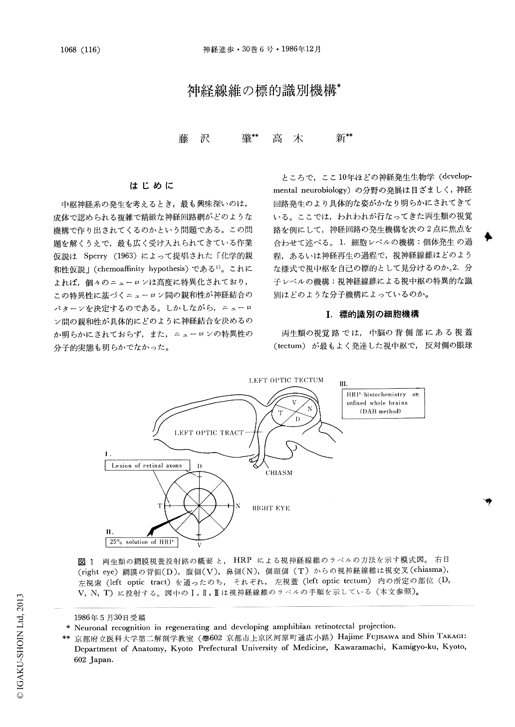
ところで,ここ10年ほどの神経発生生物学(developmental neurobiology)の分野の発展は目ざましく,神経回路発生のより具体的な姿がかなり明らかにされてきている。ここでは,われわれが行なってきた両生類の視覚路を例にして,神経回路の発生機構を次の2点に焦点を合わせて述べる。1.細胞レベルの機構:個体発生の過程,あるいは神経再生の過程で,視神経線維はどのような様式で視中枢を自己の標的として見分けるのか。2.分子レベルの機構:視神経線維による視中枢の特異的な識別はどのような分子機構によっているのか。
One of the most important biological problems to be solved is how the ordered neuronal connections in the central nervous system (CNS) are built up during regeneration or development. To clarify cellular and molecular mechanisms underlying the ordered patterns of neuronal connection in the CNS, the amphibian retinotectal projection system is a very convenient model. We analized a spatial and temporal mode of growth of retinal axons in regeneration and development, after filling retinal axons with a neuronal tracer horseradish peroxidase, and clarified cellular mechanisms related to the formation of the ordered retinotectal projection map. In regeneration of the optic nerve of adult newt Cynops pyrrhogaster, each retinal axon arrived at their correct target regions within the tectum (a major visual center in amphibians) at 6 th week after optic nerve transection. Ariving of retinal axons at correct targets was a result of preferential selection of appropriate branches of individual axons. In development of the visual systems in Xenopus tadpoles, newly formed retinal axons with a growth cone at each growing tip were retinotopically aligned within the tectum. Branching of retinal axons started when the tips of retinal axons had arrived at their approximate sites of innervation within the tectum, and preferential selection of appropriate branches was followed. Axonal sprouting and selection of appropriate branches continued throughout larval life, until the growth of the retina and tectum had completely stopped. These anatomical findings indicate that direct interaction between retinal axons and visual centers are primarily important to establish specific neuronal connection, at least in the initial steps of the retinotectal connection. To clarify molecular background of the cellular interaction between retinal axons and visual centers, we are screening target specific marker molecules by the monoclonal antibody producing technique. We have obtained a monoclonal antibody (MAb A5) which strongly bind to the visual centers of the Xenopus tadpole, and a monoclonal antibody (MAb B2) which widely bind to the brain tissues but not to the visual centers. Immunohistochemical and immunochemical properties of A5 antigen suggest that this antigen may be a candidate of optic nerve target marker molecules.
Copyright © 1986, Igaku-Shoin Ltd. All rights reserved.