Japanese
English
- 有料閲覧
- Abstract 文献概要
- 1ページ目 Look Inside
I.はじめに
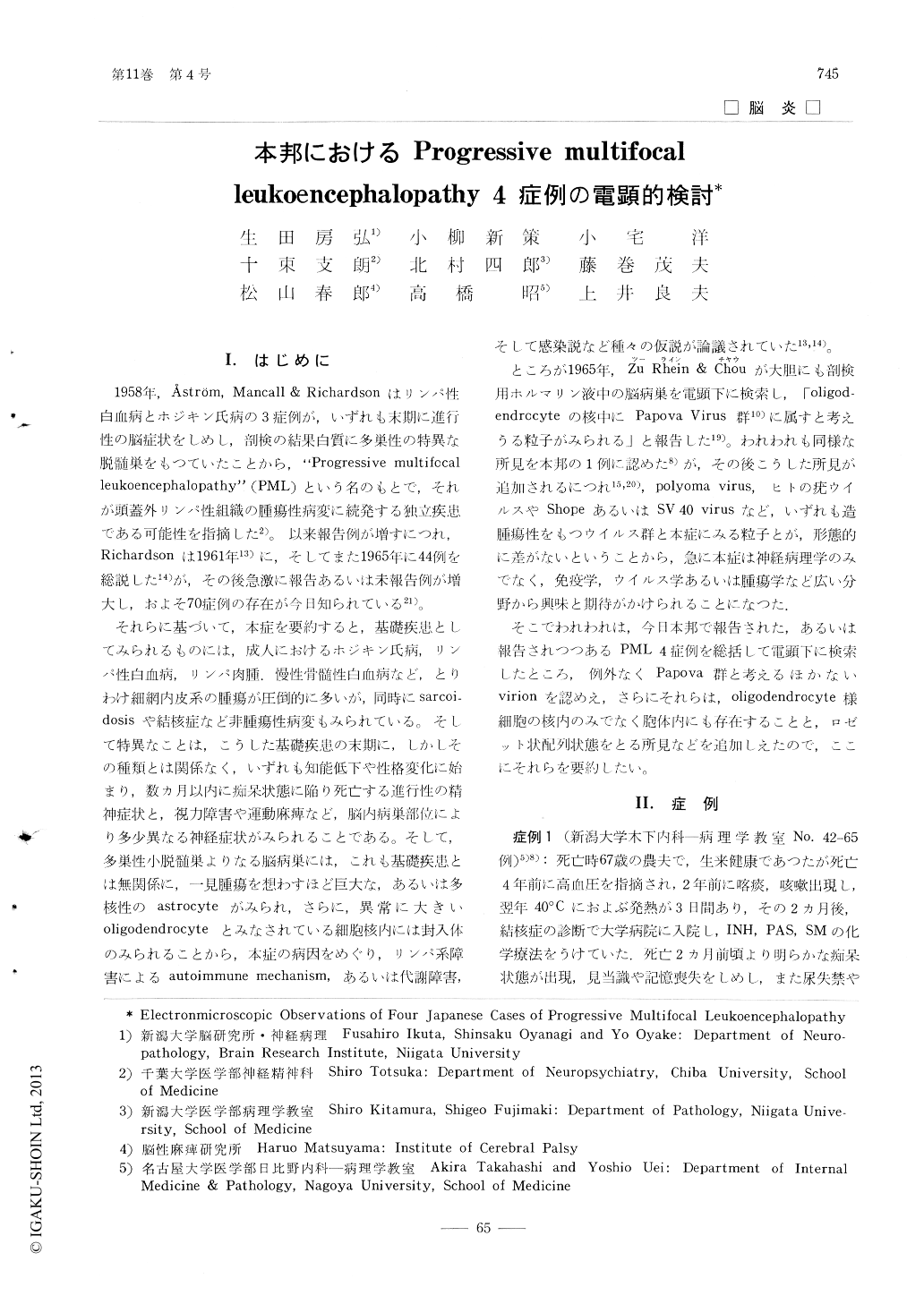
1958年,Åström,Mancall & Richardsonはリンパ性白血病とポジキン氏病の3症例が,いずれも末期に進行性の脳症状をしめし,剖検の結果白質に多巣性の特異な脱髄巣をもつていたことから,"Progressive multifocalleukoencephalopathy"(PML)という名のもとで,それが頭蓋外リンパ性組織の腫瘍性病変に続発する独立疾患である可能性を指摘した2)。以来報告例が増すにつれ,Richardsonは1961年13)に,そしてまた1965年に44例を総説した14)が,その後急激に報告あるいは未報告例が増大し,およそ70症例の存在が今日知られている21)。
それらに基づいて,本症を要約すると,基礎疾患としてみられるものには,成人におけるホジキン氏病,リンパ性白血病,リンパ肉腫,慢性骨髄性白血病など,とりわけ細網内皮系の腫瘍が圧倒的に多いが,同時にsarcoidosisや結核症など非腫瘍性病変もみられている。そして特異なことは,こうした基礎疾患の末期に,しかしその種類とは関係なく,いずれも知能低下や性格変化に始まり,数カ月以内に痴呆状態に陥り死亡する進行性の精神症状と,視力障害や運動麻痺など,脳内病巣部位により多少異なる神経症状がみられることである。
The following four autopsy cases of progressive multifocal leukoencephalopathy were studied by electron microscope. The first case was 67 year old male who suffered lung fuberculosis and interstitual fibrosis for two years. The second case of 37 year old female expired of reticulum cell sarcoma she suffered for ten months. The third case of 70 year old male revealed lungtuberculosis. The last case was a man who ceased at the age of 41 after 8 years' history of Hodgkin's sarcoma. Their terminal mental deterioration and neurological signs were observed for about 2 months, 3 months, 50 days and 5 months respectively. All four brain lesions demonstrated electron dense spherical virions, approximately 40mμ in diameter. Most of them were located among the widely dispersed nuclear chromatin and a few were detected in the cytoplasma of large round cells, which were presumed to be the altered oligodendrocytes in the lesion. Filamentous forms of the slightly smaller diamter were almost always found interspersed among the spherical virions. The latter virions often demonstrated several groups of their unique crystalline arrays. Each virion located in their central zones exhibited a central electron lucent spot in crossed form, but the virions in peripheral zones were noted to be more parenchymatous. The certain regularities detected in such arrangements of the virion which were arranged parallel with the radial lines, hexagonal and pentagonal, starting from a point, suggested that those crystalline arrangements probably represented the morphological way of their multiplication on the diagonal lines of the virion. Beside the crystalline array, a rosette-like arrangements, combined with filamentous forms, were also noted in the nucleus.

Copyright © 1967, Igaku-Shoin Ltd. All rights reserved.


