Japanese
English
- 有料閲覧
- Abstract 文献概要
- 1ページ目 Look Inside
I.はじめに
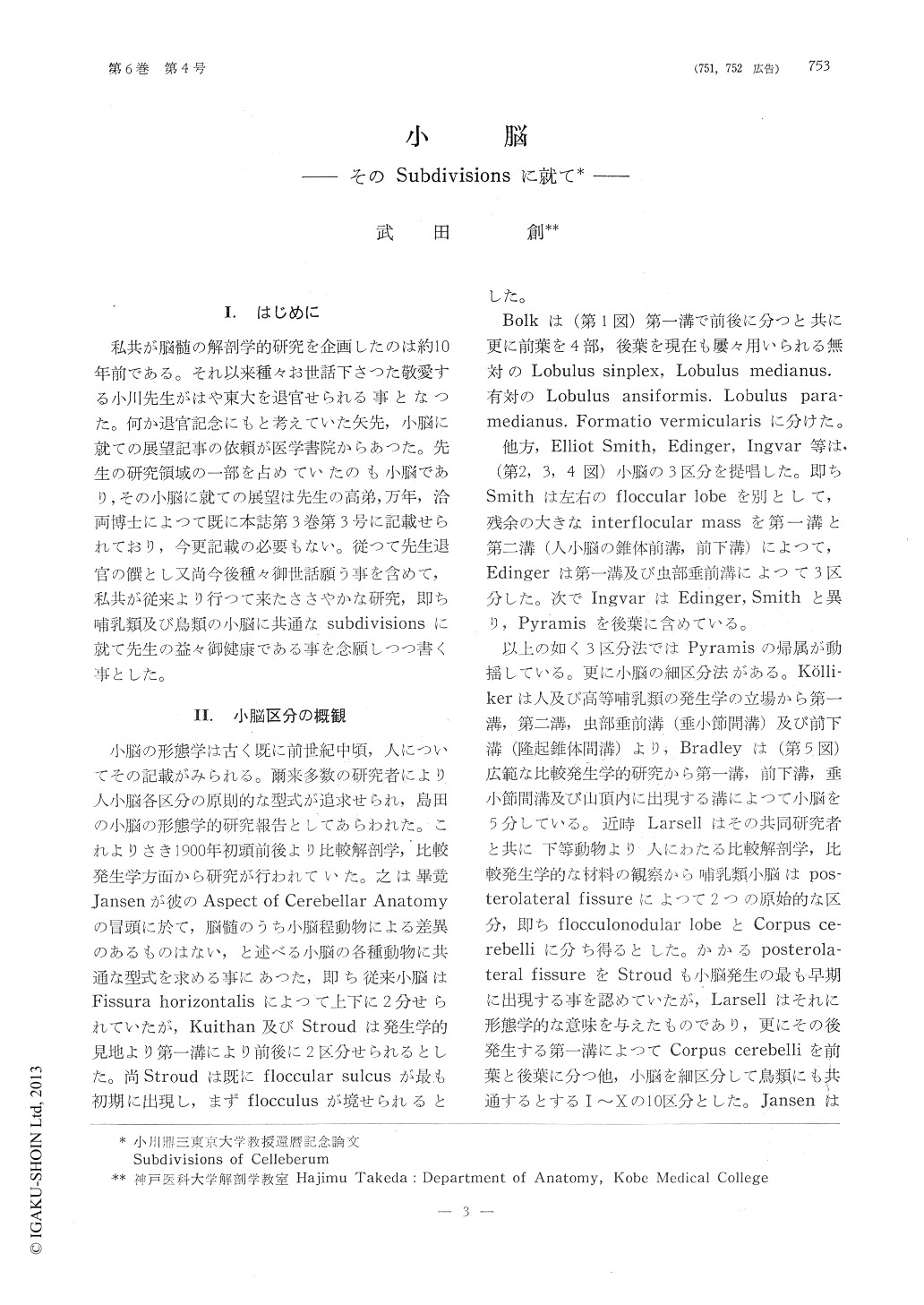
私共が脳髄の解剖学的研究を企画したのは約10年前である。それ以来種々お世話下さつた敬愛する小川先生がはや東大を退官せられる事となつた。何か退官記念にもと考えていた矢先,小脳に就ての展望記事の依頼が医学書院からあつた。先生の研究領域の一部を占めていたのも小脳であり,その小脳に就ての展望は先生の高弟,万年,洽両博士によつて既に本誌第3巻第3号に記載せられており,今更記載の必要もない。従つて先生退官の饌とし又尚今後種々御世話願う事を含めて,私共が従来より行つて来たささやかな研究,即ち哺乳類及び鳥類の小脳に共通なsubdivisionsに就て先生の益々御健康である事を念願しつつ書く事とした。
As for the divisions of cerebellar surface, there are many works since long before. However, there is no definite and final divisions today and the works which were done before are the divisions from the surface fissures.
Therefore, we tried to reconsider the divisions of the cerebellar surface from the new stand point, namely the branching of the medullary laminae by using various kinds of animals and we made out new divisions of the cerebellar cortex which is adoptable to all kinds of animals which we examined. We divided the cerebellar cortex into ten folia as Larsell did and we named each ten lamina as I - X following to the name of the surface folia, but our divisions are slightly different from Larsell's divisions as showing in fig 7.
The different point between Larsell and us are as follows ;
1) In the vermal portion of ayes are the divisions of IV and subbranch of VI and VII. Although Larsell includes IV in Culmen, we regards it as the caudal part of Lobulus centralis as mentioned below.
Then, we understood that there is commonly subbranches, namely VIa and VIb in VI and not in VII. Therefore, we regard VIb as Folium vermis instead of Larsell's divisions which regard Vila as Folium vermis.
There are two reasons why we did such divisions. The first one is that the precise observation of the reconstruction model showed that such divisions is most reasonable. The second one is the developmental study of the cerebellar medullary body and fissures which we did concerning uroloncha, pigeon, duck, hen and pig.
From the phylogenetic study in ayes, it is clear that I-V are bordered by the primary fissure and Folium V is divided first.
However, V is absorbed into the common branch of VI and VII, and in the other hand Folium II, III and IV become shallower in the lateral, although V is slightly expanding in the lateral portion.
Thus, only V shows peculiar condition. This is the reason why we regarded only V as Culmen and II, II and IV as Lobulus centralis.
2) In the mammalian cerebellum, there is a slight difference from ayes. V is regarded as Declive and VI as Folium vermis, because the primary fissure is before the Folium V. IV is corresponding to V in ayes and II and III regarded as Lobulus centralis. There is no difference in I, VII, VII, IX and X. I-IV are occuring directly from the central medullary body but II and III fuse together in primates and Human. V, VI and VII are branches of Ziehen's so called Lobulus impendens ; namely, V is the branches from rostal surface of Lobulus impendens, VI is the distal branch and VII is the proximal branch from its caudal surface. VIII is different by the kinds of animals. Namely, in human and primates it is occuring directly from the central medullary body but in carnivores it is a subbranch of IX and in ungulates and rodents it is one more branch of Lobulus impendens. No difference is seen IX and X.
The intereresting point is Lobulus centralis. This division is including three folia in ayes namely II, III and IV, but it decreases their number into twos, namely II and III in mamma-ls and it become only one (II+III) in human.
3) About the hemisphere of the cerebellarcortex there are many differences with the divisions which had been reported. Concerning the hemisphere of Pyramis, there are many theories but they may be summarized into two ; namely,
① It corresponds to Paraflocculus.
② It corresponds to Lobulus paramedianus.
First of all, in our observation of the rodents the crista of VIII approaches very closely to the crista of IX. However, these two cristal are clearly demarcated in all animals and form the posterior part of Lobulus paramedia-nus in the hemisphere.
In other an mats, it is very difficult to identify the connection of Pyramis and the posterior part of Lobulus paramedianus from the surface as the anterior and the middle parts of Lobulus paramedianus are developing well and pushing the posterior part of Lobulus paramedianus. However, we can identify the connection by the reconstruction models.
In cat VIII is swelling at the transitional portion to the hemisphere as it is curving caudally, and moreover in primates this swelling portion is consisted by the connection of two subbranches, namely well developed rostro-dorsal VIIH and caudo-ventral VIIHThe opinion that Pyramis connect to Paraflo-cculus in the hemisphere is not correct. This is the misunderstanding of authors who could not identify the medullary cristal connection between two, because of the close approach of Pyramis and Paraflocculus.
That VII is connecting with the anterior and middle parts of Lobulus paramedianus is very clear from the observation of reconstructions and therefore Tubur vermis has no connection with Lobulus ansiformis as Larsell said.
The middle part of the Lobulus paramedianus is homlogous with the Lobulus gracilis in human. Folium vermis has connection with only Lobulus ansiformis which has two portions, namely Crus I and Crus II. The groove which borders Lobulus ans:formis from Lobulus paramedianus is Fissura ansoparame-diana in animals and Fissura horizontalis in human.
The hemisphere of Uvula is Paraflocculus. Paraflocculus dorsalis and ventralis corresponds with IXHB and IXHA in mammals and ayes. The development of Paraflocculus ventalis is relatively poor in human, and it becomes Flocculus accessorius. However, Paraflocculus dorsalis is well developed in human, particu-larly toward the medial direction and form Tonsilla cerebelli.
Concerning the twisting of the posterior vermal portion the measuremental study was done. This phenomenon is especially prominent in carnivores and ungulates. As the twisting is particularly seen in Folium vermis and Tuber vermis, the ratio of surface dimensions in Folium vermis and Tuber vermis to the whole cerebellar surface was calculated and the animals which have such twisting showed more than 7 percent.

Copyright © 1962, Igaku-Shoin Ltd. All rights reserved.


