Japanese
English
- 有料閲覧
- Abstract 文献概要
- 1ページ目 Look Inside
はじめに
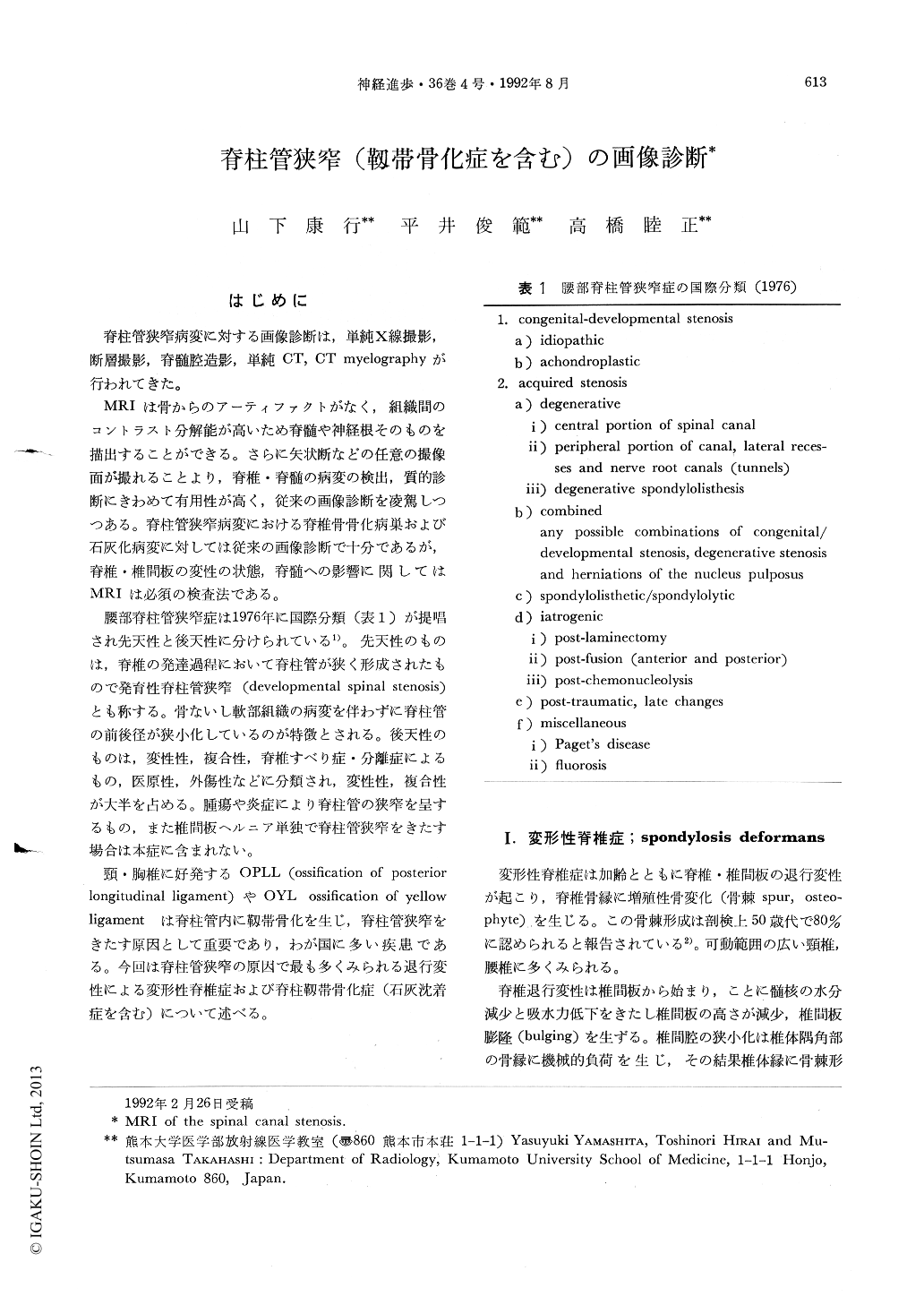
脊柱管狭窄病変に対する画豫診断は,単純X線撮影,断層撮影,脊髄腔造影,単純CT,CT myelographyが行われてきた。
MRIは骨からのアーティファクトがなく,組織間のコントラスト分解能が高いため脊髄や神経根そのものを描出することができる。さらに矢状断などの任意の撮像面が撮れることより,脊椎・脊髄の病変の検出,質的診断にきわめて有用性が高く,従来の画像診断を凌駕しつつある。脊柱管狭窄病変における脊椎骨骨化病巣および石灰化病変に対しては従来の画像診断で十分であるが,脊椎・椎間板の変性の状態,脊髄への影響に関してはMRIは必須の検査法である。
For the diagnosis of the diseases involving the spine, MRI is the procedure of choice as well as plain radiography. Spinal canal stenosis due to various etiologies is shown by MRI to better advantage. Spinal canal stenosis can be diagnosed with MRI as narrowing of the spinal canal. There is frequently associated prominent osteophyte, hypertrophy of the ligament and bulging of the disk. Osteophytes, degenerative disks, ossification of the ligament can be demonstrated as low signal intensities. The degenerated disk is shown especially well on T2 weighted images.

Copyright © 1992, Igaku-Shoin Ltd. All rights reserved.


