Japanese
English
- 有料閲覧
- Abstract 文献概要
- 1ページ目 Look Inside
I.はじめに
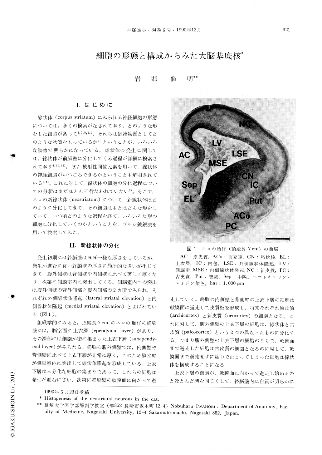
線状体(corpus striatum)にみられる神経細胞の形態については,多くの検索がなされており,どのような形をした細胞があって3,7,8,11),それらは伝達物質としてどのような物質をもっているか5)ということが,いろいろな動物で明らかになっている。線状体の発生に関しては,線状体が前脳壁に分化してくる過程が詳細に検索されており4,10,13),また放射性同位元素を用いて,線状体の神経細胞がいつごろできるかということも解明されている1,4)。これに対して,線状体の細胞の分化過程についての分析はまだほとんど行なわれていない2)。そこで,ネコの新線状体(neostriatum)について,新線状体はどのように分化してきて,その細胞はもとはどんな形をしていて,いつ頃どのような過程を経て,いろいろな形の細胞に分化していくのかということを,ゴルジ鍍銀法を用いて検索してみた。
The neostriatal neurons originated from the medial and lateral striatal elevation which protruded into the ventrolateral region of the lateral ventricle. As seen in Golgi sections obtained from a fetus with a 7 cm craniorump length, somata of the radial glia lined the ventricular surface of the striatal elevation, composing the ependymal layer. Each radial glia had a long process extending toward the pial surface. A thick subependymal layer composed of densely packed small cells was adjacent to the ventrolateral surface of the ependymal layer.

Copyright © 1990, Igaku-Shoin Ltd. All rights reserved.


