Japanese
English
- 有料閲覧
- Abstract 文献概要
- 1ページ目 Look Inside
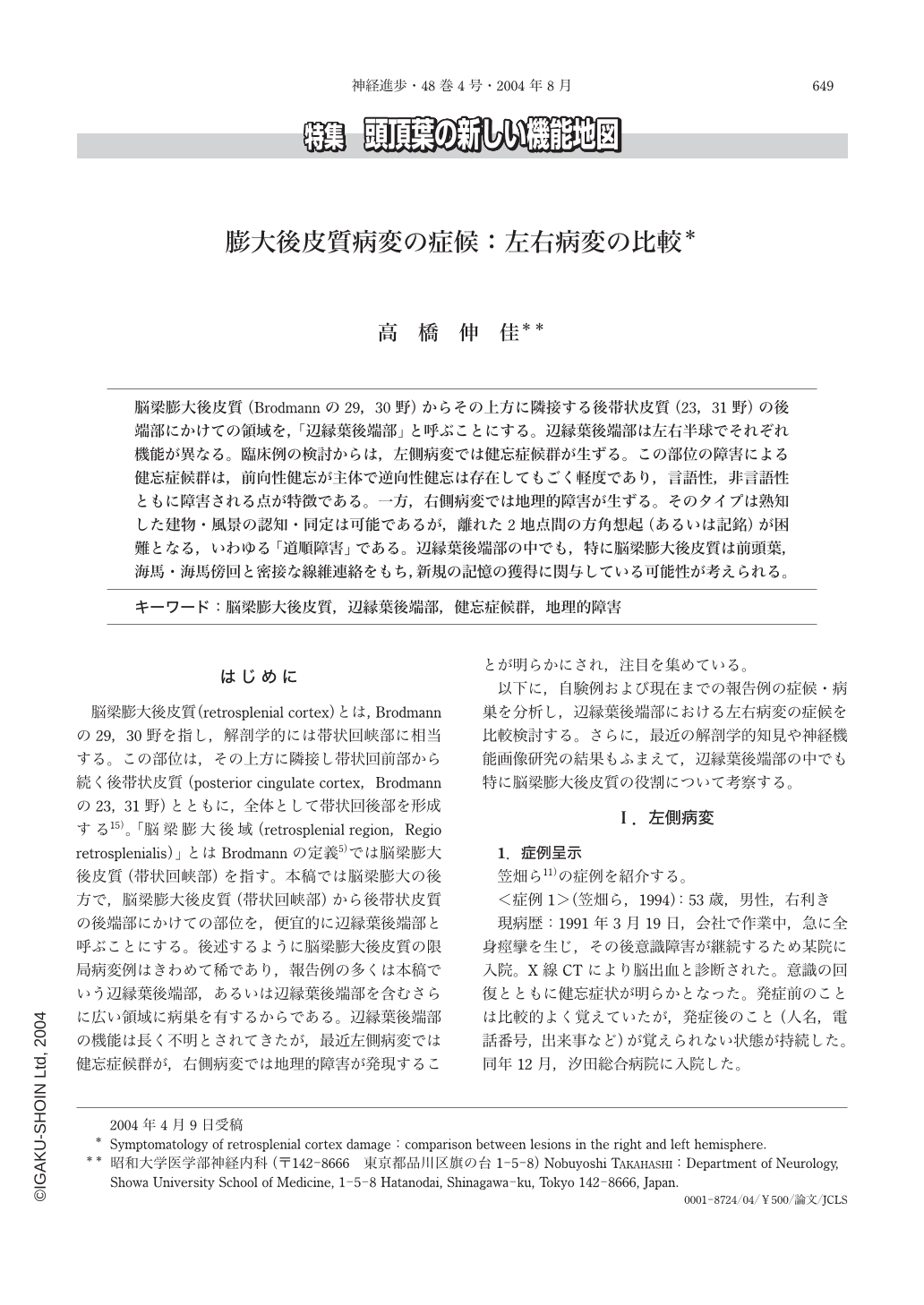
脳梁膨大後皮質(Brodmannの29,30野)からその上方に隣接する後帯状皮質(23,31野)の後端部にかけての領域を,「辺縁葉後端部」と呼ぶことにする。辺縁葉後端部は左右半球でそれぞれ機能が異なる。臨床例の検討からは,左側病変では健忘症候群が生ずる。この部位の障害による健忘症候群は,前向性健忘が主体で逆向性健忘は存在してもごく軽度であり,言語性,非言語性ともに障害される点が特徴である。一方,右側病変では地理的障害が生ずる。そのタイプは熟知した建物・風景の認知・同定は可能であるが,離れた2地点間の方角想起(あるいは記銘)が困難となる,いわゆる「道順障害」である。辺縁葉後端部の中でも,特に脳梁膨大後皮質は前頭葉,海馬・海馬傍回と密接な線維連絡をもち,新規の記憶の獲得に関与している可能性が考えられる。
The retrosplenial cortex, and the posterior cingulate cortex located in the upper part of the retrosplenial cortex, are referred to as the posterior cingulate area. For convenience, we refer to the region that is located posterior to the splenium and extends from the retrosplenial cortex to the posterior end of the posterior cingulate cortex as the posterior end of the limbic lobe.
It is known that the symptoms of the lesion of the posterior end of the limbic lobe differ between right and left hemisphere. A retrosplenial lesion in the left hemisphere is associated with amnesic syndrome. The amnesic syndrome due to this lesion is predominantly anterograde amnesia, occasionally associated with very mild retrograde amnesia, and characteristically both verbal and nonverbal memories are impaired. In contrast, a right hemisphere lesion is associated with topographical disorientation. Patients can recognize and identify familiar buildings and landscapes, but cannot remember the positional relation between two locations within a comparatively wide range of familiar surroundings.
The posterior end of the limbic lobe, especially the retrosplenial cortex, is known to be closely connected with the frontal lobe, hippocampus, and parahippocampal gyrus. Investigations carried out on patients who have lesions confined to the retrosplenial cortex and the results of recent functional neuroimaging studies suggest that the retrosplenial cortex might have a role in the encoding of novel information.

Copyright © 2004, Igaku-Shoin Ltd. All rights reserved.


