Japanese
English
- 有料閲覧
- Abstract 文献概要
- 1ページ目 Look Inside
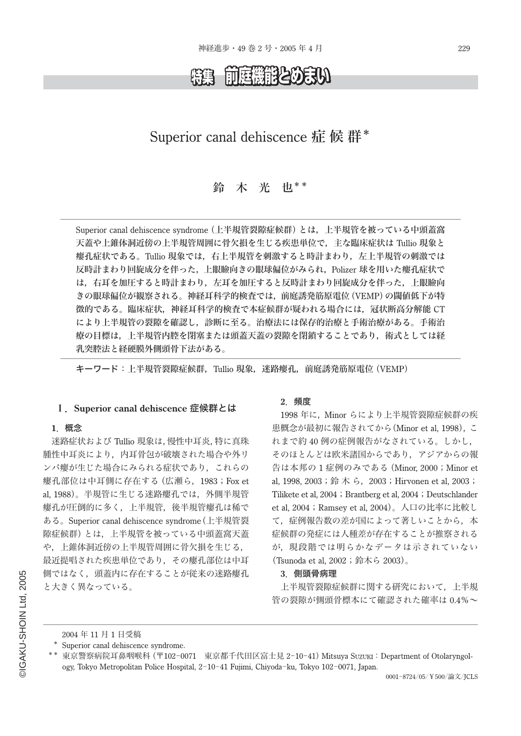
Superior canal dehiscence syndrome(上半規管裂隙症候群)とは,上半規管を被っている中頭蓋窩天蓋や上錐体洞近傍の上半規管周囲に骨欠損を生じる疾患単位で,主な臨床症状はTullio現象と瘻孔症状である。Tullio現象では,右上半規管を刺激すると時計まわり,左上半規管の刺激では反時計まわり回旋成分を伴った,上眼瞼向きの眼球偏位がみられ,Polizer球を用いた瘻孔症状では,右耳を加圧すると時計まわり,左耳を加圧すると反時計まわり回旋成分を伴った,上眼瞼向きの眼球偏位が観察される。神経耳科学的検査では,前庭誘発筋原電位(VEMP)の閾値低下が特徴的である。臨床症状,神経耳科学的検査で本症候群が疑われる場合には,冠状断高分解能CTにより上半規管の裂隙を確認し,診断に至る。治療法には保存的治療と手術治療がある。手術治療の目標は,上半規管内腔を閉塞または頭蓋天蓋の裂隙を閉鎖することであり,術式としては経乳突腔法と経硬膜外側頭骨下法がある。
Superior canal dehiscence syndrome is a recently established disease unit, which present Tullio phenomenon and fistula symptom due to the dehiscence of bone overlying the superior semicircular canal. In this syndrome, the dehiscence of bone overlying the superior semicircular canal often involves both the bilateral temporal bones. The cause of the dehiscence of the bone is unknown whether it is due to the acquired disturbance of the bone remodeling or due to congenital bone defect. Patients suffering from the superior canal dehiscence syndrome develop vertical-torsional eye movements(closely aligned with the plane of the superior semicircular canal of the affected side)with vertigo and oscillopsia in response to loud sounds or maneuvers that change middle ear or intracranial pressure such as valsalva. Valsalva with closed nostrils results in a vertical-tortional nystagmus with slow phases directed upward and outward from the ear which is suspected to be responsible for the symptoms and signs. The nystagmus evoked by valsalva with a closed glottis produces eye movement in the opposite direction. Patients occasionally experience disequilibrium, hyperacusis, gaze-evoked tinnitus and hearing loss. Usually, the caloric response of affected ear is normal. Vestibular evoked myogenic potential(VEMP) is useful for detecting excitability or hypersensitivity in the saccular macula. The threshold of a vestibular evoked myogenic potential(VEMP) is generally low in the affected ear in superior canal dehiscence syndrome. A coronal section of high-resolution computed tomography of the temporal bones is useful in identifying the dehiscence of the bone overlying the superior semicircular canal in patients suffering from the superior canal dehiscence syndrome. Canal plugging or resurfacing of the canal dehiscence by the transmastoid or extradural subtemporal approach alleviates the symptoms and signs. A plug composed of a piece of fascia and bone dust when placed in the lumen of the bony canal obliterates plug it. The plug is covered with a fascia and a bone graft harvested from the inner surface of the middle fossa bone flap. The resurfacing of the canal dehiscence using the fascia directly overlying the membranous canal is followed by a bone graft and then, placing an outer layer of fascia. The major complications of such surgeries are cochlear or vestibular dysfunction of the operated ear.

Copyright © 2005, Igaku-Shoin Ltd. All rights reserved.


