Japanese
English
- 有料閲覧
- Abstract 文献概要
- 1ページ目 Look Inside
囊胞性病変の3例
[症例1]
患者 59歳,男性.
主訴 心窩部痛.
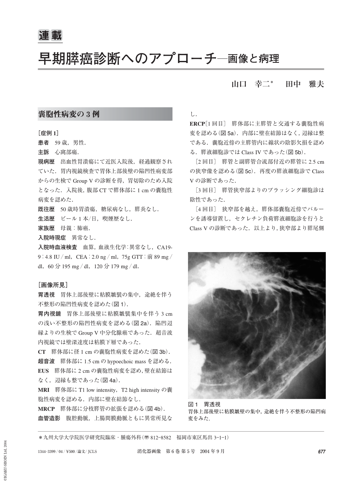
現病歴 出血性胃潰瘍にて近医入院後,経過観察されていた.胃内視鏡検査で胃体上部後壁の陥凹性病変部からの生検でGroup Vの診断を得,胃切除のため入院となった.入院後,腹部CTで膵体部に1 cmの囊胞性病変を認めた.
(Case 1)
A 59-year-old man was referred to us for the treatment of early gastric cancer. Computed tomography incidentally found a cystic lesion, measuring 1 cm, in the body of the pancreas. Endoscopic retrograde pancreatography showd stensosis of the main pancreatic duct in the neck of the pancreas and a cystic lesion in the body of the pancreas. Cytology of the pancreatic juice from the main pancreatic duct distal to the stenosis showed malignant cells. Computed tomography and magnetic resonance imagings showed no mass lesion other than the cystic lesion. Distal pancreatectomy was done with partial gastrectomy. Histological examination showed a well differentiated adenocarcinoma with minimal invasion and intraductal papillary mucinous adenoma with mild dysplasia. Early gastric carcinoma was also evident, measuring 1.5 cm. He is doing well seven years after the operation.
(Case 2)
A 55-year-old man was diagnosed as having chronic active hepatitis and diabetes mellitus ten years ago and as having hepatocellular carcinoma three years ago. Follow-up computed tomography showed a cystic lesion, measuring 2.5 cm, in the tail of the pancreas. Endoscopic retrograde pancreatography showed a cystic lesion in the tail and cytology of the pancreatic juice showed atypical cells highly suggestive malignancy. Distal pancreatectomy and splenectomy was done. Histological examination showed a carcinoma in-situ in the branch duct apart from intraductal papillary mucinous adenoma with mild dysplasia. He died of pancreatic carcinoma in the remnant pancreas seven years after the operation.
(Case 3)
A 63-year-old man had endoscopic mucosal resection for rectal carcinoma. Follow-up computed tomography showed a multilocular cystic lesion in the uncinate portion of the pancreas. Endoscopic retrograde pancreatography showed a stenosis of the main pancreatic duct at the body as well as a cystic lesion in the uncinate process of the pancreas. Brushing cytology of the stenosis of the main pancreatic duct showed malignant cells. Ultrasonography, magnetic resonance imagings and endoscopic ultrasonography showed only a cystic lesion the uncinate process. Total pancreatectomy was done for the cystic lesion and the stenosis. Histological examination showed two small invasive ductal carcinomas and intraductal mucinous adenoma of the pancreas with mild dysplasia. He is doing well five years and ten months after the operation.
(Shokakigazo 2004 ; 6 : 677―686)
Copyright © 2004, Igaku-Shoin Ltd. All rights reserved.