Japanese
English
- 有料閲覧
- Abstract 文献概要
- 1ページ目 Look Inside
- 参考文献 Reference
はじめに
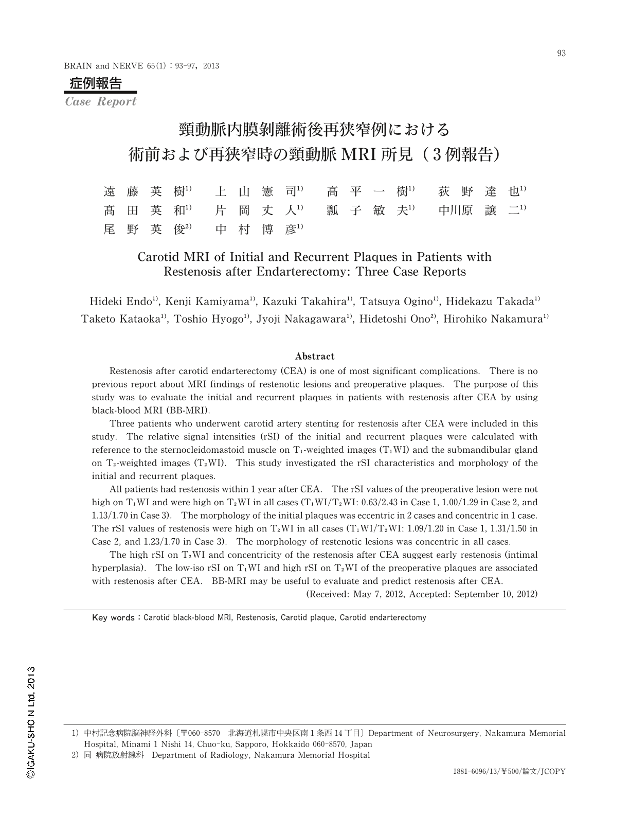
頸部頸動脈狭窄症の評価において,頸動脈MRI(magnetic resonance imaging)の有用性が多数報告されている1-4)。しかしながら,術後再狭窄例に関する報告はない。頸動脈内膜剝離術(carotid endarterectomy:CEA)後再狭窄に対して頸動脈ステント留置術(carotid artery stenting:CAS)を施行した3症例において,術前病変および再狭窄病変の頸動脈MRI所見(信号強度パターン,形態)を評価・検討したので報告する。
Abstract
Restenosis after carotid endarterectomy (CEA) is one of most significant complications. There is no previous report about MRI findings of restenotic lesions and preoperative plaques. The purpose of this study was to evaluate the initial and recurrent plaques in patients with restenosis after CEA by using black-blood MRI (BB-MRI).
Three patients who underwent carotid artery stenting for restenosis after CEA were included in this study. The relative signal intensities (rSI) of the initial and recurrent plaques were calculated with reference to the sternocleidomastoid muscle on T1-weighted images (T1WI) and the submandibular gland on T2-weighted images (T2WI). This study investigated the rSI characteristics and morphology of the initial and recurrent plaques.
All patients had restenosis within 1 year after CEA. The rSI values of the preoperative lesion were not high on T1WI and were high on T2WI in all cases (T1WI/T2WI: 0.63/2.43 in Case 1, 1.00/1.29 in Case 2, and 1.13/1.70 in Case 3). The morphology of the initial plaques was eccentric in 2 cases and concentric in 1 case. The rSI values of restenosis were high on T2WI in all cases (T1WI/T2WI: 1.09/1.20 in Case 1, 1.31/1.50 in Case 2, and 1.23/1.70 in Case 3). The morphology of restenotic lesions was concentric in all cases.
The high rSI on T2WI and concentricity of the restenosis after CEA suggest early restenosis (intimal hyperplasia). The low-iso rSI on T1WI and high rSI on T2WI of the preoperative plaques are associated with restenosis after CEA. BB-MRI may be useful to evaluate and predict restenosis after CEA.
(Received: May 7, 2012, Accepted: September 10, 2012)

Copyright © 2013, Igaku-Shoin Ltd. All rights reserved.


