Japanese
English
- 有料閲覧
- Abstract 文献概要
- 1ページ目 Look Inside
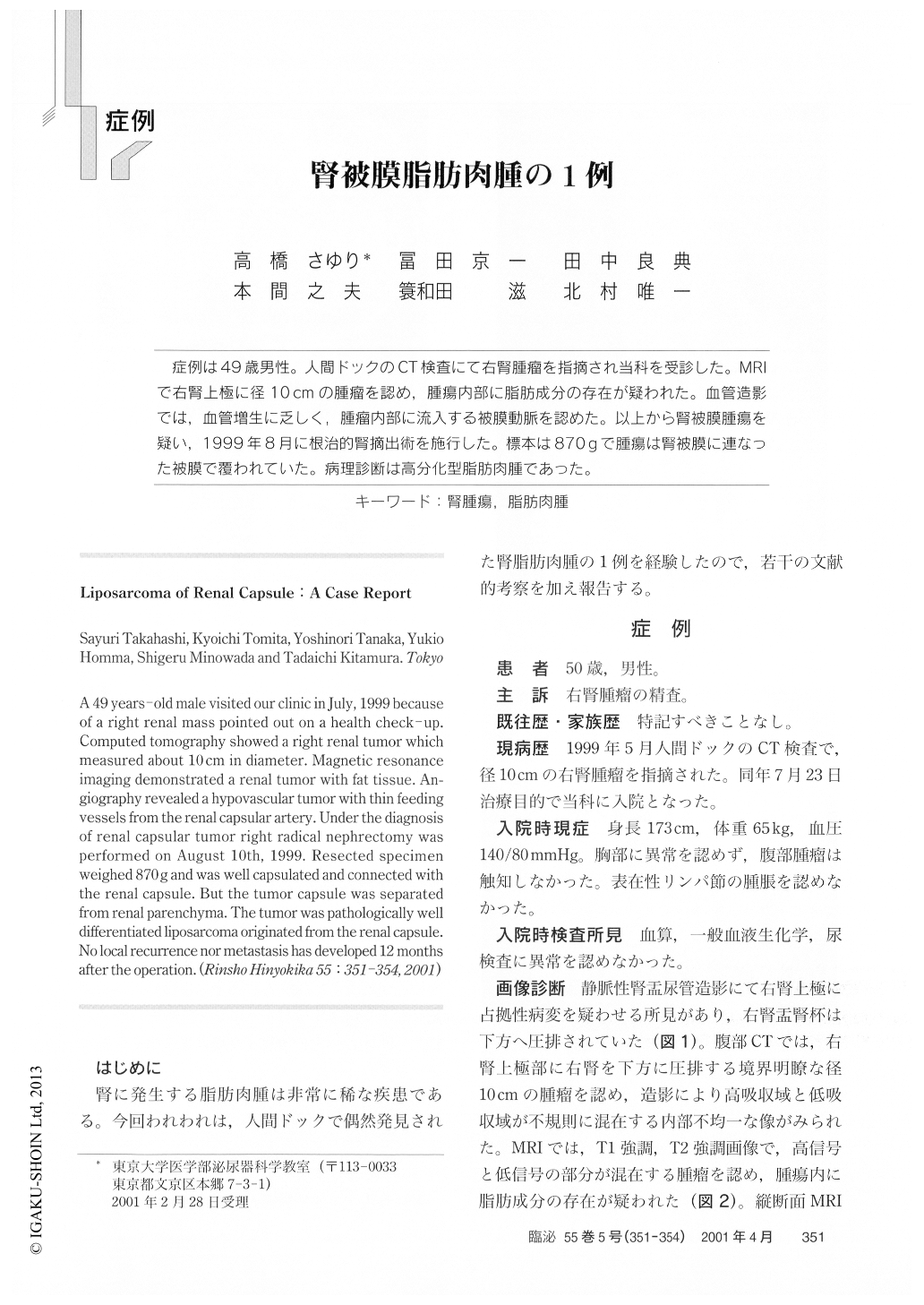
症例は49歳男性。人間ドックのCT検査にて右腎腫瘤を指摘され当科を受診した。MRIで右腎上極に径10cmの腫瘤を認め,腫瘍内部に脂肪成分の存在が疑われた。血管造影では,血管増生に乏しく,腫瘤内部に流入する被膜動脈を認めた。以上から腎被膜腫瘍を疑い,1999年8月に根治的腎摘出術を施行した。標本は870gで腫瘍は腎被膜に連なった被膜で覆われていた。病理診断は高分化型脂肪肉腫であった。
A 49 years-old male visited our clinic in July, 1999 because of a right renal mass pointed out on a health check - up. Computed tomography showed a right renal tumor which measured about 10 cm in diameter. Magnetic resonance imaging demonstrated a renal tumor with fat tissue. Angiography revealed a hypovascular tumor with thin feeding vessels from the renal capsular artery. Under the diagnosis of renal capsular tumor right radical nephrectomy was performed on August 10th, 1999. Resected specimen weighed 870g and was well capsulated and connected with the renal capsule. But the tumor capsule was separated from renal parenchyma.

Copyright © 2001, Igaku-Shoin Ltd. All rights reserved.


