- 有料閲覧
- 文献概要
- 1ページ目
血管由来の悪性腫瘍は,はなはだまれな疾患である。血管を構成する内皮細胞から由来した悪性腫瘍が,malignant hemangioendothelioma悪性血管内皮細胞腫であり,一般に本腫瘍の好発部位は,顔面,頭部で,老人に多くみられ1,2),肺,肝,骨などに転移して急速に死の転帰をとる3)。もちろん,若年者にもみられるし4),頭部,顔面以外の部位にも原発する5)。血管に由来する悪性腫瘍がまれであるためか,その術語には,統一を欠く。たとえば,malignant hemangioendoth-eliomaを,angiosarcomaと同義語とするもの2,5)と,両者を別のentityの疾患名として用いるもの6)とがある。
血管由来の悪性腫瘍としては,hemangioend-othelioma-angiosarcomaと異なるものとして,malignant hemangiopericytomaがあるが,この概念はかなり把握し難い。またKaposiの肉腫は,その臨床所見,経過,組織学的所見にhem-angioendothelioma-angiosarcomaと異なる点があるにせよ7),また腫瘍細胞が未分化であり,分化傾向の特徴をつかみ難い場合には,鑑別診断が困難となり,そこに諸説の唱えられる余地が生ずる。
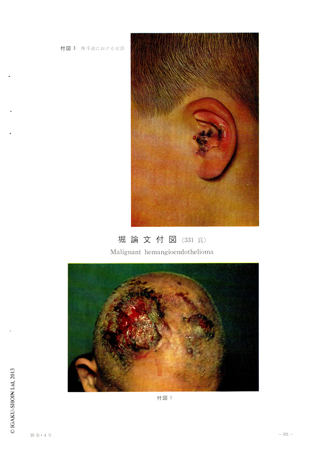
A 60-year-old man of this disease with a red, soft, hemorrhagic tumor on the diffuse, red macule on the frontal and parietal region was reported.
On autopsy, the local infiltration of the tumor down into the bone marrow of the skull, and the metastasis to the right pleura, right lung and 6th vertebra were revealed.
Histologic specimen of the skin tumor showed the dilatation of the vessels, proliferation and atypicality of the vascular endothelial cells, edema and bleeding in the cutis. The endothelial cells proliferated toward the inside and outside of the vessels. Mytosis of the endothelial cells and multinucleated endothelial cells were also proved.
Electron microscopic pictures showed enlarged endothelial cells, rich in organelles of the cyto-plasma, and with atypical nuclei. The lumen which surrounded these endothelial cells was narrow or stenotic.
These histologic and electron microscopic findings suggested that this disease was a tumor malignantly transformed from the previously existed endothelial cells.

Copyright © 1971, Igaku-Shoin Ltd. All rights reserved.


