Japanese
English
- 有料閲覧
- Abstract 文献概要
- 1ページ目 Look Inside
はじめに
スポロトリクム症は近年増加の傾向にあり,関東地方を中心に各地から多数の報告に接するが,その大部分は顔面または上肢に生じたものであり,下肢を侵した症例は非常にまれである。われわれは今回,右足背および右下腿に生じた皮膚リンパ管型の本症の1例を経験したので報告する。
A 66-year-old farmer's wife near Tokyo suffered from a shoe sore on the back of the right foot in August, 1963. It was treated by a doctor with ointments, roentgen, etc, but 9 lesions developed upward gradually as if it spread along the lymphatic vessels.
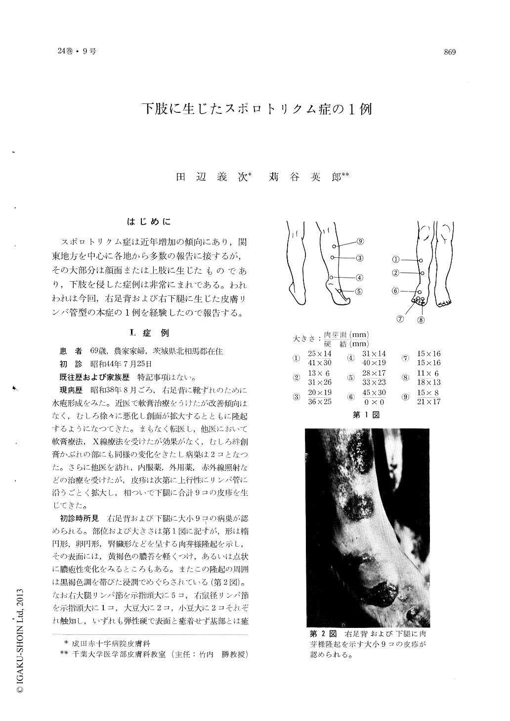
At her first visit on June 25, 1969, the lesions from 6×13 mm to 30×45 mm in size, were oval or kidney-shaped, granulomatous tumors surrounded with dark-brown infiltrated areas.
There were 5 lymph nodes, 1 cm in diameter, in the right femoral region and several lym-ph nodes, up to 1 cm in diameter, in the right inguinal region.
Histologic specimen showed pseudoepitheliomatous hyperplasia and a diffuse chronic nonspecific granulomatous infiltrate composed of neutrophils (microabscesses in places), epithelioid cells, giant cells, lymphocytes and plasma cells. PAS stain reveled free spores, spores in giant cells and asteroid bodies. Sporotrichum schenckii was identified by slide culture. This fungus sho-wed yeast-like colonies in the brain-heart infusion agar with 0.5% glucose at 37℃. Thus, two phases of this fungus was proved.
Peroral administration of potassium iodide cured the lesions clinically, histologically and mycologically.

Copyright © 1970, Igaku-Shoin Ltd. All rights reserved.


