Japanese
English
- 有料閲覧
- Abstract 文献概要
- 1ページ目 Look Inside
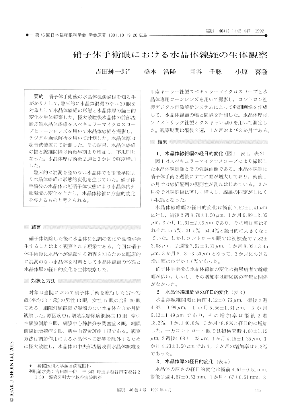
硝子体手術後の水晶体混濁過程を知る手がかりとして,臨床的に水晶体混濁のない30眼を対象として水晶体線維の形態と水晶体厚の経日的変化を生体観察した。極大散瞳後水晶体の前部浅層皮質水晶体線維をスペキュラーマイクロスコープとコーンレンズを川いて水晶体線維を撮影し,デジタル画像解析を川いて計測した。水晶体厚は超音波装置にて計測した,その結果,水晶体線維の幅と線維間隔は術後早期より増加し,不規則となった。水晶体厚は術後2週と3か月で軽度増加した。
臨床的に混濁を認めない水晶体でも術後早期より水晶体線維に形態的変化を生じていた。硝子体手術後の水晶体は無硝子体状態により水晶体内外部環境の変化をきたし,水晶体線維に形態的変化を与えるものと考えられる。
We evaluated the lens fibers and lens thickness in 30 eyes before and after vitreous surgery. The series included proliferative diabetic retinopathy 10 eyes, tractional retinal detachment 9 eyes, central retinal vein occlusion 8 eyes among others. The crystalline lens was clear in all the eyes prior to surgery. We photographed the lens fibers in the anterior cortex using specular microscope and cone lens. The lens thickness was measured by ultrasono-graphy. We observed the width of lens fibers and fiber spaces to increase and to become irregular from the early postoperative stage on. The thick-ness of the lens increased slightly during the post-operative 2 weeks to 3 months. The observed changes in lens morphology seemed to suggest that the barrier in the lens cortex was destroyed by the absence of anterior vitreous after vitreous surgery.

Copyright © 1992, Igaku-Shoin Ltd. All rights reserved.


