Japanese
English
- 有料閲覧
- Abstract 文献概要
- 1ページ目 Look Inside
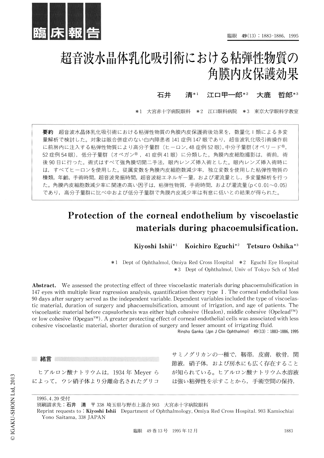
超音波水晶体乳化吸引術における粘弾性物質の角膜内皮保護術後効果を,数量化1類による多変量解析で検討した。対象は眼合併症のない白内障患者141症例147眼であり,超音波乳化吸引術操作前に前房内に注入する粘弾性物質により高分子量群(ヒーロン,48症例52眼),中分子量群(オペリード®,52症例54眼),低分子量群(オペガン®,41症例41眼)に分類した。角膜内皮細胞撮影は,術前,術後90日に行った。術式はすべて強角膜切開二手法,眼内レンズ挿入術とした。眼内レンズ挿入術時には,すべてヒーロンを使用した。従属変数を角膜内皮細胞数減少率,独立変数を使用した粘弾性物質の種類,年齢,手術時間,超音波発振時間,超音波総エネルギー量,および灌流量とし,多変量解析を行った。角膜内皮細胞数減少率に関連の高い因子は,粘弾性物質,手術時間,および灌流量(p<0.01〜0.05)であり,高分子量群に比べ中および低分子量群で角膜内皮減少率は有意に低いとの結果が得られた。
We assessed the protecting effect of three viscoelastic materials during phacoemulsification in 147 eyes with multiple liear regression analysis, quantification theory type I . The corneal endothelial loss 90 days after surgery served as the independent variable. Dependent variables included the type of viscoelas-tic material, duration of surgery and phacoemulsification, amount of irrigation, and age of patients. The viscoelastic material before capsulorhexis was either high cohesive (Healon), middle cohesive (OpeleadTM)or low cohesive (OpeganTM). A greater protecting effect of corneal endothelial cells was associated with less cohesive viscoelastic material, shorter duration of surgery and lesser amount of irrigating fluid.

Copyright © 1995, Igaku-Shoin Ltd. All rights reserved.


