Japanese
English
- 有料閲覧
- Abstract 文献概要
- 1ページ目 Look Inside
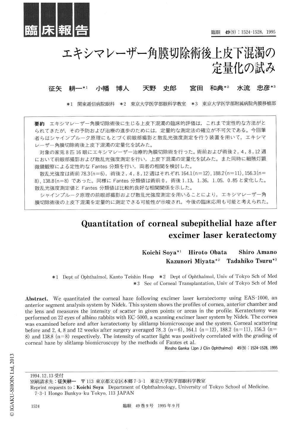
エキシマレーザー角膜切除術後に生じる上皮下混濁の臨床的評価は,これまで定性的な方法がとられてきたが,その予防および治療の進歩のためには,定量的な測定法の確立が不可欠である。今回筆者らはシャインプルーク原理にもとづく前眼部撮影と散乱光強度測定を行う装置を用いて,エキシマレーザー角膜切除術後上皮下混濁の定量化を試みた。
対象の家兎8匹16眼にエキシマレーザー治療的角膜切除術を行った。術前および術後2,4,8,12週において前眼部撮影および散乱光強度測定を行い,上皮下混濁の定量化を試みた。また同時に細隙灯顕微鏡観察による定性的なFantes分類を行い,両者の相関を検討した。
散乱光強度は術前78.3(n=6),術後2,4,8,12週はそれぞれ164.1(n=12),188.2(n=11),156.3(n=8),138.8(n=8)であった。同様にFantes分類値は術前0,術後1.13,1.36,1.05,0.85と変化した。散乱光強度測定値とFantes分類値は比較的良好な相関関係を示した。
シャインプルーク原理の前眼部撮影および散乱光強度測定を用いることにより,エキシマレーザー角膜切除術後の上皮下混濁を定量的に測定できる可能性が示唆され,今後の臨床応用も可能と考えられた。
We quantitated the corneal haze following excimer laser keratectomy using EAS-1000, an anterior segment analysis system by Nidek. This system shows the profiles of cornea, anterior chamber and the lens and measures the intensity of scatter in given points or areas in the profile. Keratectomy was performed on 22 eyes of albino rabbits with EC-5000, a scanning excimer laser system by Nidek. The cornea was examined before and after keratectomy by slitlamp biomicroscope and the system. Corneal scattering before and 2, 4, 8 and 12 weeks after surgery averaged 78.3 (n=6), 164.1 (n=12), 188.2 (n=11), 156.3 (n= 8) and 138.8 (n=8) respectively. The intensity of scatter light was positively correlated with the grading of corneal haze by slitlamp biomicroscopy by the methods of Fantes et al.

Copyright © 1995, Igaku-Shoin Ltd. All rights reserved.


