Japanese
English
- 有料閲覧
- Abstract 文献概要
- 1ページ目 Look Inside
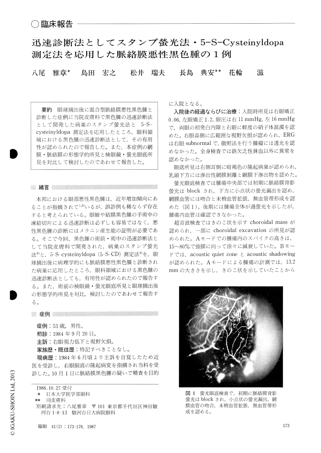
眼球摘出後に混合型脈絡膜悪性黒色腫と診断した症例に当院皮膚科で黒色腫の迅速診断法として開発した病巣のスタンプ螢光法と5-S-cysteinyldopa測定法を応用したところ,眼科領域における黒色腫の迅速診断法として,その有用性が認められたので報告した.また,本症例の網膜・脈絡膜の形態学的所見と検眼鏡・螢光眼底所見を対比して検討したのであわせて報告した.
A new diagnostic method for malignant melanoma is described. It consists of two parts : stamp fluorescence method and quantitative determination of 5-S-cysteinyl-dopa (5-S-CD). Each method usually takes 30 minutes. Choroidal melanoma cells emit intenst specific fluores-cence, while pigmented nevus cells and pigment basal epithelioma cells emit weak or no fluorescence. The specific fluorescence emitted by melanotic melanoma cells is mainly due to the presence of 5-S-CD.
We applied this method to a 53-year-old male, who manifested a mushroom-shaped brown tumor in the temporal sector in his right fundus. The tumor mea-sured 15 × 15 × 12 mm. It proved, histologically, to have ruptured the Bruch's membrane to invade the inner retina. It was diagnosed, histopathologically, as mixed-type malignant melanoma of the choroid, composed spindle A, spindle B and epithelioid cells. The amount of 5-S-CD in the melanoma tissue was 87 ng/mg, while the amount of 5-S-CD the pigmented nevus and pigmented basal epithelioma tissues varied from 1.3 to 37.9 ng/mg.
These results indicate that the stampt fluorescence and quantitative determination of 5-S-CD are useful methods for rapid diagnosis. They promise to be of value in needle biopsy of intraocular melanoma as well as biopsy of extraocular melanoma prior to surgical treatment.
Rinsho Ganka (Jpn J Clin Ophthalmol) 41(2) : 173-178, 1987
Copyright © 1987, Igaku-Shoin Ltd. All rights reserved.