Japanese
English
- 有料閲覧
- Abstract 文献概要
- 1ページ目 Look Inside
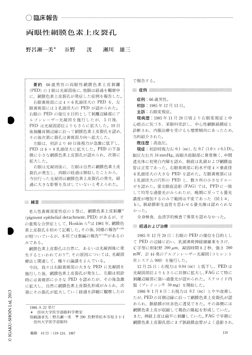
66歳男性の両眼性網膜色素上皮剥離(PED)の1眼は光凝固後に,他眼は経過を観察中に,網膜色素上皮裂孔が発症した症例を報告した.
右眼黄斑部には4×6乳頭径大のPEDを,左眼黄斑部には1乳頭径大のPEDが認められた.右眼のPEDの復位を目的として剥離辺縁部にアルゴンレーザー光凝固を施行したが,5日後,PEDは光凝固部位よりもさらに拡大した.19日後剥離耳側辺縁に沿って網膜色素上皮裂孔を認め,その後次第に裂孔は黄斑部方向へ拡大した.
左眼は,初診より40日後視力が急激に低下し,PEDは6×8乳頭径大に拡大した.PEDの下鼻側に小さな網膜色素上皮裂孔が認められ,次第に拡大した.
右眼は光凝固後に,左眼は自然に網膜色素上皮裂孔が発生し,両眼の経過は類似したことから,今回行った光凝固は網膜色素上皮裂孔の発生,経過に大きな影響を及ぼしていないと考えられた.
A 66-year-old male presented with serous detach-ment of the retinal pigment epithelium located in the macula in both his eyes. The size of the detachment was, approximately, 4 × 6 disc diameters (DD) in the right eye and 1 DD in the left.
The right eye was treated by placing a row of argon laser photocoagulation along the margin of the detach-ment avoiding the papillomacular bundle. The serous detachment started to enlarge along the coagulated site 5 days after the treatment. A tear of the retinal pigment epithelium was formed at the temporal edge of the detachment 19 days later. The remaining detached retinal pigment epithelium retracted nasally forming curled folds.
The patient noted sudden decline in vision in his left eye 40 days after the initial examination. The detach-ment of retinal pigment epithelium had enlarged to about 6x 8 DD in size. A tear formed at the nasal edge of the detachment which gradually enlarged.
Because of the similarity in the clinical course of the tear in the retinal pigment epithelium, photocoagulationdid not appear to have induced tear of the retinal pigment epithelium.
Rinsho Ganka (Jpn J Clin Ophthalmol) 41(2) : 119-124, 1987

Copyright © 1987, Igaku-Shoin Ltd. All rights reserved.


