Japanese
English
- 有料閲覧
- Abstract 文献概要
- 1ページ目 Look Inside
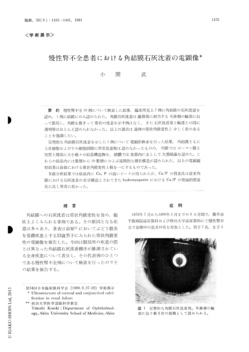
慢性腎不全10例について検索した結果,臨床所見上7例に角結膜の石灰沈着を認め,1例に結膜にのみ認められた。角膜石灰沈着は瞼裂部に相当する耳鼻側の輪部に沿って限局し,角膜を横ぎって帯状の沈着を示す例はなく,また石灰沈着部と輪部との間に透明帯はほとんど認められなかった。以上の諸点は通例の帯状角膜変性と少しく差のあることを強調したい。
定型的な角結膜石灰沈着を示した1例について電顕的検索を行った結果,角結膜ともに上皮細胞およびその細胞間隙に異常沈着物は認めなかったものの,角膜ではボーマン膜と実質上層部に大小種々の結晶構造物を,結膜では実質内に主として大型結晶を認めた。これらの結晶内には数層から10数層におよぶ規則的な層状構造が認められた。以上の電顕観察結果は前報における帯状角膜変性と軌を一にするものであった。
X線分析結果では結晶内にCa,Pの高いピークが得られたが,Ca/Pの質量比は従来角膜における石灰沈着の化学構造とされてきたhydroxyapatiteにおけるCa/Pの理論的質量比に比し異常に低かつた。
A serial examination of 10 cases with chronic renal failure revealed the presence of corneal and conjunctival calcification in 7 cases and of con-junctival one in 1 case. Corneal calcification ap-peared as an opaque semilunar arch inner to the limbus and in the interpalpebral area. The calcifi-cation did not extend across the whole cornea as in band shaped degeneration. There was no clear zone between the corneal opacity and the limbus. Corneal calcification appeared as granules scatter-ed under the epithelium in the interpalpebral fissure.

Copyright © 1981, Igaku-Shoin Ltd. All rights reserved.


